JOMI Thematic Abstract Review: May/June 2019

Reading time: 4 minutes

jomi_banner

Understanding the Role of Titanium Particles and Corrosion in Peri-implantitis

Peri-implantitis, from its recognition as an emerging concern in implant care, has been encased in confusion and controversy. Questions remain as to the treatment approaches, prevalence, incidence, and etiology. This thematic review of the literature suggests the etiology of peri-implantitis is founded on two main concepts. One concept is focused on the microbial challenge to the host-implant interface. This is a well-established etiologic model of a microbially induced inflammatory response consistent with periodontitis. The other etiologic concept looks at the role of titanium surface alterations and the release of titanium particles into the tissues as initiators of an inflammatory response. This latter model is consistent with orthopedic osteolysis. A review of our more recent literature begins to merge these two pathogenic concepts.

A systematic review by Delgado-Ruiz and Romanos (2018) describes several possible sources for titanium particles to enter the tissues as well as a role for corrosion to contribute to the development of an inflammatory response over the lifetime of the implant. Mombelli et al (2018) offer a critical review of the role of titanium particles and biocorrosion in the development of the inflammatory lesion. Their analysis suggests that while there is clear evidence that titanium particles are associated with an inflammatory response, it is postulated that the titanium particles may be a consequence of the inflammation rather than the cause of the inflammation. That is, this inflammatory environment may contribute to the biocorrosion of the implant surface.

In contrast, Noronha et al (2018) evaluated recent literature regarding the potential for titanium particles to stimulate an inflammatory response. Their findings are consistent with the titanium particles contributing to an inflammatory response, and suggest an important role for macrophages and neutrophil phagocytosis of the particles in this inflammatory response. Further, this report suggests that there may be an important relationship between the length of time of exposure to the titanium particles and the degree of inflammatory response. This time-dependence is consistent with our epidemiologic data showing an increasing prevalence of peri-implantitis over 5 or more years following placement.

Eger et al (2018) discuss their work using an animal model to show that titanium particles induce an inflammatory response, including resultant bone loss. This study also went a step further by using blockers to specific inflammatory cytokines. These inflammatory blockers were able to inhibit inflammation and bone loss in the presence of the titanium particles, therefore identifying a specific role for inflammatory cytokines in the development of titanium particle–induced peri-implantitis. Similarly, Wang et al (2019) used a different animal model to show that titanium particles can create inflammation and bone loss. This study took a different approach to inhibit the inflammatory response by blocking macrophage activity, but similar to Eger et al (2018), they found that the inflammation-induced bone loss could be inhibited. Most interestingly, this study also found that the titanium particles were able to induce inflammatory bone loss in the absence of bacterial infection.

Lastly, Daubert et al (2018) presented a unifying model that suggests titanium particles may play a role in triggering inflammation by modifying the peri-implant microbiome and that there is an interaction between the microbiome and titanium particles and corrosion. This very interesting study found that the submarginal microbial profile was more closely associated with the presence of titanium particles than with the presence or absence of peri-implantitis. This study suggests that the microbial environment and the titanium-induced inflammatory responses contribute through a codependent phenomenon to an increased susceptibility for peri-implantitis with our patients. Taken together, it appears that peri-implantitis is a complex, multifactorial condition developing through pathologic mechanisms triggered by both microbial and material interactions with the host. Going forward, our successful management and prevention of peri-implantitis will likely become increasingly guided by understanding the relationships of each of these relevant factors.

Thomas W. Oates, DMD, PhD
University of Maryland
Baltimore, Maryland, USA


Thematic Abstract Review Section Editor
Clark M. Stanford, DDS, PhD
The University of Illinois at Chicago, Chicago, Illinois, USA


Abstracts referenced:

Daubert D, Pozhitkov A, McLean J, Kotsakis G. Titanium as a modifier of the peri-implant microbiome structure. Clin Implant Dent Relat Res 2018;20:945–953.

Noronha Oliveira M, Schunemann WVH, Mathew MT, et al. Can degradation products released from dental implants affect peri-implant tissues? J Periodontal Res 2018;53:1–11.

Mombelli A, Hashim D, Cionca N. What is the impact of titanium particles and biocorrosion on implant survival and complications? A critical review. Clin Oral Implants Res 2018;29(suppl 18):s37–s53.

Delgado-Ruiz R, Romanos G. Potential causes of titanium particle and ion release in implant dentistry: A systematic review. Int J Mol Sci 2018;19(11).

Eger M, Hiram-Bab S, Liron T, et al. Mechanism and prevention of titanium particle-induced inflammation and osteolysis. Front Immunol 2018;9:2963.

Wang X, Li Y, Feng Y, Cheng H, Li D. Macrophage polarization in aseptic bone resorption around dental implants induced by Ti particles in a murine model. J Periodontal Res 2019. [Epub ahead of print].


This article was originally published in the May/June 2019 edition (Vol 34, issue 3) of The International Journal of Oral & Maxillofacial Implants.

©2019 BY QUINTESSENCE PUBLISHING CO, INC. PRINTING OF THIS DOCUMENT IS RESTRICTED TO PERSONAL USE ONLY. NO PART MAY BE REPRODUCED OR TRANSMITTED IN ANY FORM WITHOUT WRITTEN PERMISSION FROM THE PUBLISHER.

Posted in Research, The International Journal of Oral & Maxillofacial Implants, Thematic Abstract Review | Leave a comment

Challenging Scientific Paradigms: Zero Bone Loss Concepts

Reading time: 8 minutes

With today’s scientific advancements, it’s hard to appreciate the hygiene practices that help us avoid life-threatening illnesses. We forget that for the majority of human history, lifespans weren’t nearly as long as they are now. It’s equally difficult to imagine how people once believed foul odors, evil spirits, astrological arrangements, and bathing could cause disease epidemics. Bloodletting, the popular treatment practice used to purge the “bad blood” believed to cause infection, is simply horrifying to think about. These ideas may sound outrageous today, but people in the Middle Ages accepted them just as fervently as we rely on our hand sanitizers, personal hygiene, and food and waste safety measures of the 21st century.

Continually testing and challenging these scientific paradigms is a cornerstone of scientific advancement.

In the 1800s, germ theory replaced the centuries-old miasma theory of dangerous air clouds. Thanks to the discoveries of John Snow, Louis Pasteur, and Robert Koch, we now know that microscopic organisms—bacteria and viruses—are the true causes of illness. Armed with this knowledge, we have developed ways to treat and prevent their invasion into the body, thereby contributing to longer, healthier lives.

A hundred years from now, people may be just as quick to dismiss germ theory in favor of new scientific breakthroughs, but that’s the beauty of progress. Continually testing and challenging these scientific paradigms is a cornerstone of scientific advancement. Dr Tomas Linkevičius, DDS, Dip Pros, PhD, set out to do just that with Zero Bone Loss Concepts, his revolutionary new book on implant dentistry. His research has uncovered breakthrough treatment practices to prevent bone loss, has examined the multitude of factors that contribute to it, and has determined that multidisciplinary clinicians are uniquely positioned to treat it.

Zero Bone Loss

“The concept that bone loss is unavoidable should be abandoned.”

Crestal bone loss has accompanied implant treatment for so long that it has historically been considered the natural result. In fact, this “norm” has been codified. When Dr Linkevičius was a postgraduate resident of prosthodontics, almost 15 years ago, he was met with this existing paradigm.

“More experienced colleagues explained to me that this bone loss is considered to be unavoidable and urged me to just accept this fact,” Dr Linkevičius recalls. “However, for me, it was obvious that there must be reasons for it and that those factors should be researched and explained.”

Different reactions of crestal bone level to dental implants. (a) Zero bone loss. (b) Stable remodeling. (c) Progressive bone loss. (d) Bone growth.

(a and b) Crestal bone stability 7 years after augmentation and placement of implants without platform switching or a conical connection. The 7-year follow-up shows bone growth and intensive corticalization of bone.

He and his team began digging deeper into the interworkings of unavoidable bone loss and soon made discoveries that proved otherwise. Their results dared to challenge the existing paradigm, dismissing the old standards of implant dentistry where 1 mm of bone loss is considered normal.

“The concept that bone loss is unavoidable should be abandoned,” Dr Linkevičius explains.

Zero bone loss, defined as long-term crestal bone stability, plays an important role in implant function. Bone stability is one key factor toward a good esthetic outcome, leading to patient satisfaction. Dr Linkevičius’s research stresses that the goal should always be prevention of bone loss, especially in two major situations that require bone levels be as stable as possible: implants in the esthetic zone and the use of short implants. The shorter the implant, the more dangerous the same amount of bone loss.

Remineralization of crestal bone around implants (V3, MIS). (a) Delivery day. (b and c) After 1 year.

Dr Linkevičius and his team have developed four ways to increase vertical soft tissue thickness, which is one of the critical surgical factors for ensuring zero bone loss. These are outlined in the book with a chapter devoted to each method. The text also looks a step further than bone loss prevention—there could even be remineralization or bone growth.

Different methods of vertical soft tissue augmentation. (a and b) Flattening of the alveolar ridge. (c and d) Subcrestal implant placement. (e and f) Tent-pole technique. (g and h) Vertical augmentation.

Crestal Bone Loss: A Multifactorial Issue

Now that the concept and cause of bone loss can be better understood through the text and through Dr Linkevičius’s worldwide lectures, a common focus of readers and attendees is to pinpoint a single cause.

“Crestal bone loss is a multifactorial issue, and there is no single most important factor.”

“When I hear lecturers talking about the most important factor, or audience members asking to name one, I immediately perceive a lack of deep understanding of the subject,” Dr Linkevičius says. “I want readers to understand that crestal bone loss is a multifactorial issue, and there is no single most important factor.”

To demonstrate this in the text, he describes an apple metaphor.

“Imagine a basket of apples,” he says. “Each apple represents a separate factor that influences crestal bone stability. The purpose of research is to take one apple out of the basket and study it alone, eliminating other confounding factors. Clinical studies must be designed so that the factor in question—the single apple—can be studied as objectively as possible. The difficult part is that after research is complete, the apple must be returned to the basket, meaning that in clinical reality all factors operate simultaneously.”

Basket of apples metaphor

The Value of the Multidisciplinary Clinician

Dr Linkevičius was trained as a prosthodontist, and for the first 5 years of his professional career, his practice remained limited to prosthetic work. This seemed to work for a while, until he hit a wall.

“I soon realized that without the proper surgical knowledge and techniques, I simply could not deliver the results that my patients deserved,” Dr Linkevičius says. “I strongly feel that whenever possible, it is ideal for all treatment to be performed by the same specialist who possesses knowledge of implant design, biology, and prosthetics and can therefore achieve zero bone loss around every implant.”

“Respecting surgical factors is simply not enough. We need to take prosthetic factors into consideration as well.”

To meet the needs of his patients, he set out to widen his skillset, adding “implant dentist” to his résumé. Subsequently, his implant research has benefitted from his personal/professional journey, and his book reflects those professional practices.

“If we want to have crestal bone stability after restoration of implants, respecting surgical factors is simply not enough,” Dr Linkevičius says. “We need to take prosthetic factors into consideration as well—they are equally important.”

Put another way, “A surgeon without prosthetic knowledge and a prosthodontist lacking surgical awareness may never accomplish the most desired results, as both parts are equally responsible for bone stability.”

From establishing crestal bone stability through surgical factors to maintaining that stability through prosthetic factors, the book combines surgical and prosthetic advice, making it a unique, one-stop resource of clinical procedures backed by solid clinical evidence.

Impossible Is Nothing

Dr Linkevičius’s goal for Zero Bone Loss Concepts is that clinicians thoroughly understand and can practically apply the zero bone loss breakthrough, multifactorial concept, and multidisciplinary value. However, he is all too aware that his research into implant dentistry has only touched the tip of the iceberg on crestal bone loss. While his book presents the latest on the concept to date, the scientific paradigm he has set in motion—away from beliefs of the past 30 years and toward new research—will continue to change and shift over time. The process of compiling his research into this book has intimately shown him how scientific research is constantly evolving in a never-ending cycle. Case in point: New information was being generated while the book was being printed, which will inform future editions.

Until then, Dr Linkevičius stands by a favorite saying that “impossible is nothing” when it comes to improving crestal bone loss levels around implants. He encourages clinicians to discover this for themselves by applying the concepts detailed in the book.

“The definition of ‘best’ treatment is not permanent,” he stresses.

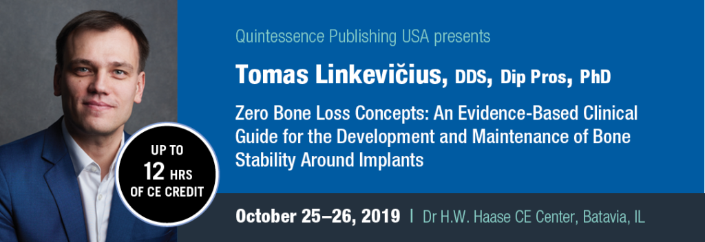
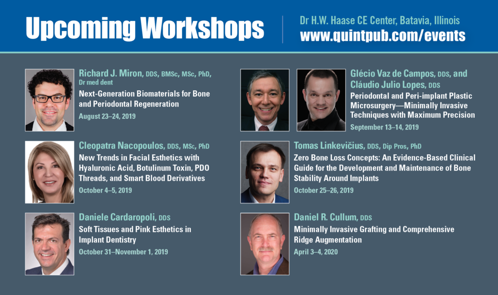
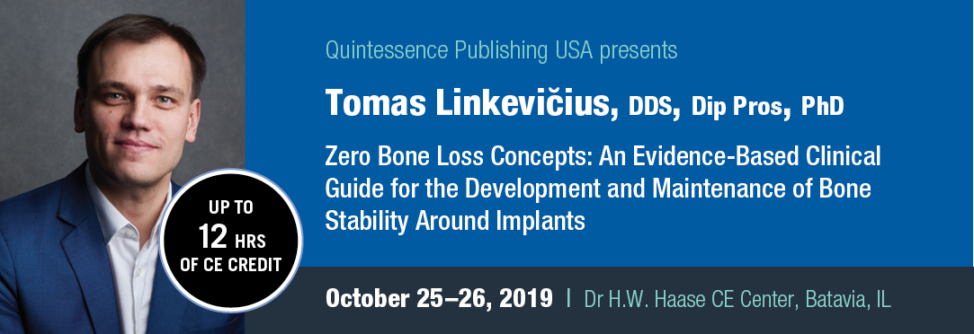
He is determined to continue making new versions of that “best” treatment possible for practicing clinicians for years to come. Clinicians may get an inside look at the latest treatment practices in October at his upcoming workshop at the Dr H.W. Haase CE Center in Batavia, IL.


Tomas Linkevičius, DDS, Dip Pros, PhD, is a professor at Vilnius University in Vilnius, Lithuania, where he completed his postgraduate studies in prosthetic dentistry and obtained his prosthodontist specialty. Dr Linkevičius received his dental degree in 2000 from Kaunas Medical University in Kaunas, Lithuania, and defended his doctoral dissertation at Riīga Stradins University in Rīga, Latvia, in 2009. Dr Linkevičius has authored dozens of publications in international peer-reviewed journals. He has also contributed to several books, including Cementation in Dental Implantology (Springer, 2015) and Implants in the Aesthetic Zone (Springer, 2019). During the course of his research, Dr Linkevičius developed zero bone loss concepts, an evidence-based clinical protocol that demonstrates how to achieve and maintain crestal bone stability around dental implants. He lectures internationally on this topic and is an active member of several professional organizations, such as the Academy of Osseointegration and the European Academy of Osseointegration. Dr Linkevičius maintains a practice specializing in prosthodontics and implant dentistry and continues to study zero bone loss concepts at the Vilnius Research Group.


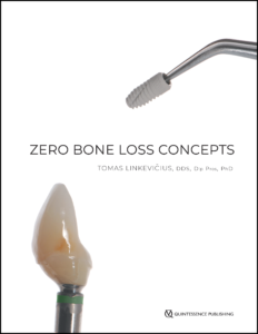
Zero Bone Loss Concepts
Tomas Linkevičius

Bone loss is still a major issue that dentists encounter, but it is a complication that can be prevented. By combining clinical experience with peer-reviewed scientific evidence, the author of this book has put together a guide that any implant specialist will find invaluable to prevent bone loss in their patients. Different strategies are presented that can be used to achieve zero bone loss years after treatment. Because successful treatment depends on both the surgical and prosthetic components, the book is divided into two parts, each focusing in depth on what must be done in each phase to promote bone stability. Case presentations detail many types of clinical situations, implant choices, and prosthetic solutions, all backed by evidence-based clinical studies that have proven success.

304 pp; 1,231 illus; ©2019; ISBN 978-0-86715-799-4 (B7994); US $198

Contents
Developing Crestal Bone Stability • Implant Design • Implant Depth • Vertical Soft Tissue Thickness • Subcrestal Implant Placement • Flattening Bone • Tent-Pole Technique • Vertical Soft Tissue Augmentation • Attached Tissues • Cement- and Screw-Retained Restorations • Individual Abutments • Fixed Partial Dentures • Angulated Abutments • Emergence Profile • Prosthetic Materials • Subgingival Materials • Supragingival Materials


This article was written by Sarah Mondello, Quintessence Publishing.

©2019 BY QUINTESSENCE PUBLISHING CO, INC. PRINTING OF THIS DOCUMENT IS RESTRICTED TO PERSONAL USE ONLY. NO PART MAY BE REPRODUCED OR TRANSMITTED IN ANY FORM WITHOUT WRITTEN PERMISSION FROM THE PUBLISHER.

Posted in Books, Feature, Implant Dentistry, Multidisciplinary, Prosthodontics | Tagged , , , , , | Leave a comment

Quintessence Roundup: June 2019

Reading time: 4 minutes

Monthly Special


Critical Thinking: Understanding and Evaluating Dental Research, Second Edition
Donald Maxwell Brunette

The second edition of this classic textbook prepares readers to identify the information they need, read it from a critical vantage point, and assess the soundness of the conclusions. It introduces issues of logic, statistics, measurement, research design, and argument. Read more

324 pp (softcover); 90 illus; ©2007; ISBN 978-0-86715-426-9 (B4269); $48 Special price! $5


Now Available


LIT: The Simple Protocol for Dental Photography in the Age of Social Media
Miguel A. Ortiz

This book is geared toward practitioners who want to master dental photography and use their photography to build their social media presence. Written for visual learners, this book breaks down the fundamentals of dental photography for clinical practice by outlining the concepts, equipment, and key protocols for taking predictable clinical photographs. The author also provides fresh insight into the ever-changing world of digital marketing and explains what you need to know to use your photography to reach your patient market on social media. Read more

248 pp; 357 illus; ©2019; ISBN 978-0-86715-802-1 (B8021); $148 Special preorder price! $118
Available later this month

Read more about LIT: The Simple Protocol for Dental Photography in the Age of Social Media here:

Dental Photography Proficiency: A Standard of Care Issue


Zero Bone Loss Concepts
Tomas Linkevičius

Bone loss is still a major issue that dentists encounter, but it is a complication that can be prevented. This book combines years of clinical experience with peer-reviewed scientific evidence to present the different strategies that can be used to achieve zero bone loss—not just months after prosthetic delivery but years after treatment. The book is divided into sections on surgical techniques and prosthetic techniques, and there are case studies with recommendations and procedures for many types of clinical situations, implant choices, and prosthetic solutions. Read more

304 pp; 1,231 illus; ©2019; ISBN 978-0-86715-799-4 (B7994); $198 Special preorder price! $158
Available later this month


Implant Therapy: Clinical Approaches and Evidence of Success, Second Edition
Edited by Myron Nevins and Hom-Lay Wang

The second edition of this volume brings together the knowledge of the foremost leaders in implant dentistry, covering all aspects of the treatment process, from decision-making and treatment planning through imaging, surgical techniques, bone and soft tissue augmentation, multidisciplinary approaches, loading protocols, and finally strategies for preventing and treating complications and peri-implantitis as well as providing effective implant maintenance therapy. This book is an indispensable resource for clinicians seeking to provide implant treatment at the highest standard of care. Read more

544 pp; 1,782 illus; ©2019; ISBN 978-0-86715-798-7 (B7987); $328 Special preorder price! $265
Available later this month


Microsurgical Endodontics
Bertrand Khayat and Guillaume Jouanny

This book provides concrete and practical information for endodontists who want to implement surgical techniques in order for them to approach their first cases with the knowledge they need. Written by two specialists in the discipline, this book outlines predictable surgical strategies and presents more than a hundred clinical cases through each stage of treatment. Read more

246 pp; 391 illus; ©2019; ISBN 978-2-36615-057-5 (BF014); US $225


Journal Highlights


jomi_banner

JOMI May/June 2019 (Vol 34, issue 3)
Issue Highlights

Featured Article—Implant Science: Cell Phone Radiation Effect on Bone-to-Implant Osseointegration: A Preliminary Histologic Evaluation in Rabbits
Kavyashree Mohan, Pindipapanahalli Venkatarayappa Harish, Sunil Kumar Mishra, and Ramesh Chowdhary

Thematic Abstract Review: Understanding the Role of Titanium Particles and Corrosion in Peri-implantitis
Thomas W. Oates

Clinical Applications: Randomized Controlled Multicenter Clinical Study Evaluating Crestal Bone Level Change of Narrow-Diameter Versus Standard-Diameter Ti-Zr Implants for Single Tooth Replacement in Anterior and Premolar Region
Saba Sameeh Ghazal, Guy Huynh-Ba, Tara Aghaloo, Serge Dibart, Stuart Froum, Robert O’Neal, and David Cochran


Dental Meetings Quintessence Will Attend in June


IADR/AADR/CADR General Session: Booth #211
hosted by the International Association for Dental Research, the American Association for Dental Research, and the Canadian Association for Dental Research June 19–22 in Vancouver, Canada

Partners in Synergy: Booth #E18
hosted by Pikos and Salama June 20–22 in Orlando, Florida


Upcoming Quintessence Events


Posted in Announcement, Books, Journals, Promotions, Roundup, Special Offer, The International Journal of Oral & Maxillofacial Implants, What's New | Leave a comment

Quintessence Roundup: May

Reading time: 11 minutes

Monthly Special


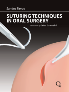
Suturing Techniques in Oral Surgery

Sandro Siervo

Proper management of soft tissues in periodontal, implant, and reconstructive surgery has become increasingly important for esthetic treatment outcomes. In order to achieve correct postoperative positioning of the soft tissues, clinicians must have a comprehensive understanding of wound healing and suturing. Drawing on a series of clinical cases, this book outlines suturing techniques for the most common surgical procedures, providing indications as well as techniques for implementation. The detailed illustrations and tables clarify difficult technical concepts, and a quick reference guide consolidates the essential information for each suture type.

240 pp; 397 illus; ©2008; ISBN 978-88-7492-120-1 (BI009); $145 Special price! $35


New Titles in Books


LIT: The Simple Protocol for Dental Photography in the Age of Social Media

Miguel A. Ortiz

In the age of digital dentistry, dental providers are under increased pressure to demonstrate proficiency in dental photography for the purposes of documentation, shade matching, and laboratory communication. Expertise in this area is fast becoming part of the standard of care and also has added value for clinicians who are looking to market themselves online. This book is geared toward practitioners who want to master dental photography and build their social media presence. Written for visual learners, this book breaks down the fundamentals of dental photography by outlining the key concepts, equipment, and lighting as well as by introducing “The Simple Protocol”—the basic day-to-day intraoral protocol that shows how easily clinical photography can be incorporated into the clinical workflow. That is where most photography books end, but this author also explores advanced techniques and demonstrates how to achieve some of the most characteristic looks in artistic dental photography, including the glossy effect, chiaroscuro, chromaticity, and texture manipulation, as well as a simple setup for taking photographs in the dental laboratory. Finally, the author provides fresh insight into the ever-changing world of digital marketing and explains what you need to know to reach your market on social media.

248 pp; 357 illus; ©2019; ISBN 978-0-86715-802-1 (B8021); $148 Special preorder price! $118
Available June 2019

Read more about LIT: The Simple Protocol for Dental Photography in the Age of Social Media here!

Dental Photography Proficiency: A Standard of Care Issue


Zero Bone Loss Concepts

Tomas Linkevičius

Bone loss is still a major issue that dentists encounter, but it is a complication that can be prevented. By combining clinical experience with peer-reviewed scientific evidence, the author of this book has put together a guide that any implant specialist will find invaluable to prevent bone loss in their patients. Different strategies are presented that can be used to achieve zero bone loss years after treatment. Because successful treatment depends on both the surgical and prosthetic components, the book is divided into two parts, each focusing in depth on what must be done in each phase to promote bone stability. Case presentations detail many types of clinical situations, implant choices, and prosthetic solutions, all backed by evidence-based clinical studies that have proven success.

304 pp; 1,231 illus; ©2019; ISBN 978-0-86715-799-4 (B7994); $198 Special preorder price! $158
Available June 2019


Implant Therapy: Clinical Approaches and Evidence of Success, Second Edition

Edited by Myron Nevins and Hom-Lay Wang

Since the publication of the first edition of this book 20 years ago, the landscape of implant dentistry has changed dramatically. Both the industry and patient demand have expanded exponentially, leaving the clinician with many decisions to make (and often as many questions to ask) regarding patient selection, surgical timing and techniques, implant types, and restorative approaches. This volume brings together the knowledge of the foremost leaders in implant dentistry, covering all aspects of the treatment process, from decision-making and treatment planning through imaging, surgical techniques, bone and soft tissue augmentation, multidisciplinary approaches, loading protocols, and finally strategies for preventing and treating complications and peri-implantitis as well as providing effective implant maintenance therapy. Filled with expert knowledge based on decades of research and clinical experience as well as abundant illustrations and clinical case presentations, this book is an indispensable resource for clinicians seeking to provide implant treatment at the highest standard of care.

544 pp; 1,782 illus; ©2019; ISBN 978-0-86715-798-7 (B7987); $328 Special preorder price! $265
Available June 2019


Microsurgical Endodontics

Bertrand Khayat and Guillaume Jouanny

When considering the correct approach to endodontic treatment, it is important to consider endodontic surgery along with conventional retreatment or tooth extraction. Not only is endodontic surgery more conservative than conventional endodontic treatment, but it can also be used to resolve difficulties that are inaccessible by conventional approach. Written by two specialists in the discipline, this book outlines predictable surgical strategies and presents more than a hundred clinical cases through each stage of treatment. This book provides concrete and practical information for endodontists who want to implement surgical techniques in order for them to approach their first cases with the knowledge they need. Also included in this book are 12 augmented reality videos, which are accessible through the “Microsurgical Endodontics” application available on the App Store (iOS) or Google Play (Android).

246 pp; 391 illus; ©2019; ISBN 978-2-36615-057-5 (BF014); US $225


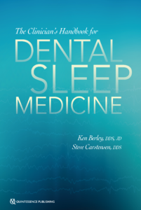
The Clinician’s Handbook for Dental Sleep Medicine

Ken Berley and Steve Carstensen

It has been estimated that 20 million Americans suffer from moderate to severe OSA, and at least one patient in five has mild OSA. The primary treatment prescribed by sleep physicians is CPAP, but patient compliance with this therapy is unacceptably low, between 25% and 50%. There is a significant opportunity for dentists to provide a viable alternative therapy—oral appliance therapy (OAT). OAT results in much better adherence to therapy than CPAP, and while OAT is not as efficacious as CPAP, this increased compliance results in comparable therapeutic results. Currently, a board-certified sleep physician is the only medical professional qualified to diagnose OSA and other sleep-related breathing disorders (SRBDs), so dentists must coordinate with a sleep physician to provide OAT. This book is the how-to guide, a gateway to a successful dental sleep medicine practice. Written by two experts in the field, it clearly delineates the dentist’s role in the treatment of SRBDs and gives practical advice for how to incorporate dental sleep medicine into an existing dental practice, not to mention how to work with sleep physicians to best support patient care. In addition to step-by-step instructions for examination, appliance selection, and follow-up care, complications of OAT, legal issues, and medical insurance and Medicare considerations are included to fully prepare the dentist for the journey into dental sleep medicine.

240 pp (softcover); 60 illus; ©2019; ISBN 978-0-86715-813-7 (B8137); Now available! $72

Read more about The Clinician’s Handbook for Dental Sleep Medicine here!

Obstructive Sleep Apnea: A Deadly Disease with a Dental Solution


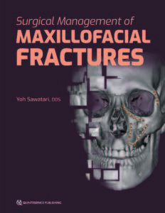
Surgical Management of Maxillofacial Fractures

Yoh Sawatari

The facial skeleton is comprised of vertical and horizontal buttresses and the intersections they create; maxillofacial fractures occur when these buttresses sustain more force than they can withstand. The objective when managing these fractures is to reverse the damage that these buttresses sustained and restore appropriate facial dimensions. Not all fractures propagate in the same pattern, so surgeons must compartmentalize the face and define the character of the individual bones. This book approaches the face one bone at a time, outlining how to evaluate each type of fracture, the indications for surgery, the surgical management, and any complications. Specific protocols for clinical, radiographic, and CT assessment are included, as well as step-by-step approaches for surgical access and internal reduction and fixation. Isolated fractures are rare with maxillofacial trauma, and the author discusses how to sequence treatment for concomitant fractures to ensure the most successful outcome. This book is a must-have for any surgeon managing maxillofacial fractures.

256 pp; 254 illus; ©2019; ISBN 978-0-86715-794-9 (B7949); Now available! $178

Read more about Surgical Management of Maxillofacial Fractures here!

Architects of the Face: Rebuilding After Maxillofacial Trauma


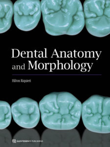
Dental Anatomy and Morphology

Hilton Riquieri

This beautiful atlas conveys not only the practical knowledge of dental anatomy but also the art of sculpting it in wax. The ideal anatomy of each dental structure is described in detail and the waxing techniques are beautifully illustrated step by step for visual reference. The author demonstrates that for every morphologic feature there is an explanation in nature, assigning significance to every minute feature of dental morphology. Organized by tooth and arch, this book views morphology through a clinical lens and repeatedly draws connections between anatomical features and clinical concepts. The fundamental knowledge presented in this text is essential for improving waxing and sculpting techniques and will be useful for students and specialists alike.

332 pp; 1,507 illus; ©2019; ISBN 978-0-86715-770-3 (B7703); Now available! $172


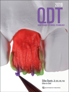
Quintessence of Dental Technology 2019: Volume 42

Edited by Sillas Duarte, Jr

QDT 2019 presents a potpourri of original articles highlighting new techniques and novel approaches for creating beautiful smiles—both in form and function. Featured articles include an innovative procedure for predictably matching a veneer to an implant crown, a program for producing a personalized smile based on its visual identity, and the Plane System for virtual functional and esthetic analysis, diagnosis, and CAD/CAM fabrication. These are but a few of the pearls found in this year’s beautifully produced annual resource for the dental technician and restorative clinician.

232 pages; 1,000+ illus; ©2019; ISBN 978-0-86715-816-8 (JQ630); Now available! $156


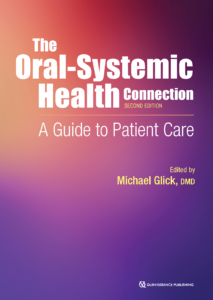
The Oral-Systemic Health Connection: A Guide to Patient Care, Second Edition

Edited by Michael Glick

As the oral-systemic health connection continues to be the focus of an overwhelming amount of scientific literature, it is important for dentists to stay informed in order to be a reliable source of information for their patients regarding both oral and overall health. This second edition of The Oral-Systemic Health Connection: A Guide to Patient Care explores the connections between oral infections and systemic diseases/conditions, incorporating feedback from scientists, practitioners, and policymakers. In addition to updated chapters about cardiovascular disease, diabetes, inflammation, and adverse pregnancy outcomes, authors have contributed new chapters about antibiotic prophylaxis, the genomic connection, common risk factors, and the economic impact of this connection. Because interpreting this research can be challenging, new chapters about causal frameworks and biostatistical assessment have been added, and specific clinical considerations for providing dental care to patients with certain conditions have been included in selected chapters. Besides providing readers with up-to-date information on the complex connection between oral and general health, this book prepares oral health care professionals to critically read and evaluate new research to ultimately benefit their patients’ overall health.

384 pp (softcover); 85 illus; ©2019; ISBN 978-0-86715-788-8 (B7888); Now available! $48


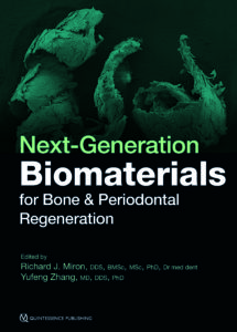
Next-Generation Biomaterials for Bone & Periodontal Regeneration

Edited by Richard J. Miron and Yufeng Zhang

New and innovative biomaterials are being discovered or created in laboratories at an unprecedented rate, but many of them remain entirely foreign to practicing clinicians. This book addresses this gap in knowledge by summarizing some of the groundbreaking research performed to date on this topic and providing case examples of these biomaterials at work. The book begins with a review of the biologic background and applications of bone grafting materials utilized in dentistry. The principles of guided tissue and bone regeneration are covered in detail, including many recent advancements in barrier membrane technologies as well as use of platelet-rich fibrin and various growth factors, and many next-generation materials that will optimize future bone and periodontal regeneration are presented. The final chapter is designed to help clinicians select appropriate biomaterials for each specific regenerative protocol. Much like one implant size and shape cannot be utilized for every indication in implant dentistry, one bone grafting material, barrier membrane, or growth factor cannot maximize regenerative outcomes in all clinical situations. This textbook teaches clinicians how to utilize biomaterials in an appropriate, predictable, and evidence-based manner.

384 pp; 960 illus; ©2019; ISBN 978-0-86715-796-3 (B7963); Now available! $218

Read more about Next-Generation Biomaterials for Bone & Periodontal Regeneration here!

Next-Generation Biomaterials: What They Are, Why You Need to Know About Them, and Which Ones Should Intrigue You


Current Issues in Journals


prd_banner

Featured article: Clinical and Histologic Evaluations of Porcine-Derived Collagen Matrix Membrane Used for Vertical Soft Tissue Augmentation: A Case Series
Algirdas Puisys, Saulius Zukauskas, Ricardas Kubilius, Mike Barbeck, Dainius Razukevičius, Laura Linkevičiene, and Tomas Linkevičius

Comparison of Three Biphasic Calcium Phosphate Block Substitutes: A Histologic and Histomorphometric Analysis in the Dog Mandible
Octavi Ortiz-Puigpelat, Basel Elnayef, Marta Satorres-Nieto, Jordi Gargallo-Albiol, and Federico Hernández-Alfaro

Immediate Loading of Implants Inserted Through Impacted Teeth in the Esthetic Area: A Series of 10 Cases with up to 7 Years of Follow-up
Francesco Amato, Ugo Macca, Giulia Amato, and Davide Mirabella

General Health Quality of Life in Patients with Temporomandibular Disorders in a Population-Based Cross-Sectional Study in Southern Brazil
Lucas B. Pigozzi, Patrícia S. Progiante, Marcos P. Pattussi, Eduardo P. Pellizzer, Patrícia K. Grossi, and Márcio L. Grossi

Intraoral Rehabilitation After Marginal Mandibulectomy
Ivana Petrovic, Jatin P. Shah, Joseph M. Huryn, Zain Uddin Ahmed, and Evan B. Rosen

Depression and Somatization in Patients with Temporomandibular Disorders in a Population-Based Cross-Sectional Study in Southern Brazil
Daniela D. S. Rehm, Patrícia S. Progiante, Marcos P. Pattussi, Eduardo P. Pellizzer, Patrícia K. Grossi, and Márcio L. Grossi

jomi_banner

Featured article: Ligature-Induced Peri-implant Bone Loss Around Loaded Zirconia and Titanium Implants
Stefan Roehling, Michael Gahlert, Simone Janner, Bo Meng, Henriette Woelfler, and David L. Cochran

Thematic abstract review: Oral Implants Made of Zirconia: An Update for 2019
Guy Huynh-Ba

TiO2-Modified Zirconia Surface Improves Epithelial Cell Attachment
Sini Riivari, Khalil Shahramian, Ilkka Kangasniemi, Jaana Willberg, and Timo O. Närhi

The Effect of Nonstrenuous Aerobic Exercise in Patients with Chronic Masticatory Myalgia
Cibele Nasri-Heir, Amey G. Patil, Olga A. Korczeniewska, Tal Zusman, Junad Khan, Gary Heir, Rafael Benoliel, and Eli Eliav

A Qualitative Study to Assess the Impact of Iatrogenic Trigeminal Nerve Injury
Sarah Barker, Tara Renton, and Susan Ormrod

Intraoral Administration of Botulinum Toxin for Continuous Dentoalveolar Neuropathic Pain: A Case Series
Isabel Moreno-Hay, Pratishtha Mishra, and Jeffrey P. Okeson

Mandibular molar distalization with clear aligners in Class III patients
Kamy Malekian, Simone Parrini, Francesco Garino, Andrea Deregibus, and Tommaso Castroflorio

Temporary Anchorage Devices (TADs) and Invisalign: A combination that can be used to meet aesthetic expectations in the orthodontic treatment of adult patients
Andrea Eliseo and Gianluigi Fiorillo

Initial therapy of occlusion
Werner Schupp, Wolfgang Boisserée, Maria Tabancis, and Julia Funke


Dental Meetings Quintessence Will Attend in May


AAOP 43rd Scientific Meeting: Booth #4
hosted by the American Academy of Orofacial Pain May 2–5 in San Diego, California

AAO Annual Session: Booth #1855
hosted by the American Association of Orthodontists May 4–7 in Los Angeles, California

CDA South: Booth #1327
hosted by the California Dental Association May 16–18 in Anaheim, California

49th JDIQ: Booth #1608–1610
hosted by the Ordre des dentistes du Québec May 24–28 in Montreal, Canada


Upcoming Quintessence Events


Posted in Announcement, Books, Journal of Oral & Facial Pain and Headache, Journals, Promotions, Roundup, Special Offer, The International Journal of Oral & Maxillofacial Implants, The International Journal of Periodontics & Restorative Dentistry, The International Journal of Prosthodontics, What's New | Leave a comment

Dental Photography Proficiency: A Standard of Care Issue

Reading time: 19 minutes

Dental photography is a hot topic right now. Facebook, Instagram, and other social media platforms, as well as dental journals and publications focused on esthetic and cosmetic dentistry, are flooded with artistic photographs where the teeth and restorations take on the role of a fashion model in a magazine spread. These photographs show dentistry as art at the highest level. But if you think this is all dental photography is and can be used for, you are mistaken. And if you think you can ignore the dental photography revolution—that it doesn’t apply to you and that it can’t improve the level of care you provide for your patients—then you are also mistaken. According to Miguel A. Ortiz, DMD, author of a new book called LIT: The Simple Protocol for Dental Photography in the Age of Social Media, dental photography provides valuable benefits to clinicians and patients alike, and more clinicians need to engage with this technology in order to elevate the level of care they provide for their patients.

“When we talk about dental photography,” Dr Ortiz says, “the first thing we need to consider is what are we trying to do for our patients. If all you’re trying to do for your patients is restore function to them via your prosthesis, then you may think that you can get away with not delivering something that looks good. But in this day and in this moment, we have the ability to make teeth that look like teeth. So if you give your patient something that functions but doesn’t look good, I think we are below the standard of care.”

“We have the ability to make teeth that look like teeth. So if you give your patient something that functions but doesn’t look good, you are below the standard of care.”

So how does dental photography impact the care that clinicians provide? Dental photography accomplishes a number of valuable goals in clinical practice. Images can be used to document stages of care, monitor the patient’s oral status over time, provide the data needed for esthetic analysis, and more. Dental photography is also one of the most effective tools for communication, whether with the patient, other specialists, or the dental laboratory. But if dental photography can be so beneficial at every stage of care—from evaluation to treatment planning, through each stage of execution, and especially during follow-up and maintenance—why do so many clinicians choose not to engage with this technology? Why are the benefits of dental photography so integral to the standard of care clinicians can provide, and how does the profession as a whole improve engagement?

The Importance of Dental Photography: Changing Perceptions

“The first barrier we have to overcome,” Dr Ortiz says, “is that of perception—the perception of what dental photography is meant for. I believe there is a generational aspect of this in which the younger generations are now more interested in topics of dental photography, and many of them for the right reasons as previously discussed. But at the same time, when we also use it for social media, the older generation of clinicians who do not engage with or see the value in social media may form the judgment that the younger clinicians are only doing dental photography for that. They may not see it as a respectable part of the profession. We need to move ahead and beyond that conception that dental photography is optional. It is only optional if you can find a better way to do what dental photography accomplishes.”

Adamo E. Notarantonio, DDS, agrees with Dr Ortiz regarding dental photography as a standard of care. “Dental photography is necessary for dentistry in every aspect,” he states. “It is important for patient communication, for records, for taking photographs of lesions and following up a week or two later to make sure nothing has changed or grown—it’s an essential part of practice. If you aren’t using dental photography, you are definitely practicing below the standard of care, without question.”

Another perception affecting the widespread adoption of dental photography is the idea that dental photography is applicable in high-end esthetic dentistry but not for general practice.

“It’s not wrong to say that the clinicians dealing with higher-end clinical cases should be a little fancier with their dental photography and knowledge,” Dr Ortiz concedes, “but my argument is that as part of the standard of care, there is a minimum that the clinician should do for each patient.”

“If you aren’t using dental photography, you are definitely practicing below the standard of care, without question.”

“Even if you’re not able to operate on the highest level, you should still be making something that looks good,” Dr Notarantonio states. “You can’t tell me that any general practitioner doing a single central incisor who sends a lab prescription that just says ‘Give me a zirconia crown, shade A1’ can’t do better by using dental photography. Without sending a photo to your ceramist, they don’t stand a chance. There’s no possible way other than sheer luck that they will get that crown right. You don’t have to practice at a fellowship level to be able to tell when there’s a monochromatic opaque crown on a tooth, and I think dental photography can help all clinicians practice at a much higher level than they can without it.”

Another barrier lies in the way dental photography is incorporated in the dental school curriculum, if it’s even incorporated at all. “The perception in dental schools right now,” Dr Ortiz explains, “is that it doesn’t need to be a major part of the curriculum. It is not being taken as seriously as the other topics covered by the curriculum. When it is taught, there is no emphasis on having the correct equipment. This kind of treatment introduces students to the perception that dental photography is optional and unimportant. We need to break that mentality because the knowledge is out there—there are no more excuses. You need a good camera just like you need a good drill, and you need to know how to use it. For the last 15 years, many of us have put the knowledge out there. We have read, we have studied, we have developed the protocols. Our colleagues don’t have to guess anymore.”

Simplifying the Concepts of Photography by Teaching More

In order to address this deficit, Dr Ortiz has organized his knowledge into a highly successful and sought-after hands-on dental photography course in an attempt to bring his colleagues up to speed. In a single day, he teaches participants the fundamentals of photography as well as intraoral and extraoral protocols through guided hands-on practice. And now, he has packaged that information plus more into one complete book: LIT: The Simple Protocol for Dental Photography in the Age of Social Media. His Simple Protocol makes the seemingly complicated possible even for true beginners. He expertly distills the high-level technical principles of photography into simplified explanations and instructions, starting with what he refers to as The Big 5: exposure, aperture, shutter speed, depth of field, and white balance.

“The Big 5 refers to the five main concepts that are most important in dental photography,” Dr Ortiz explains. “These 5 concepts will empower you to have complete control over your photographic results. Learn the Big 5 and you will be the boss of Manual Mode. Drop Auto Mode forever. You paint your own picture, not the camera.”

“Drop Auto Mode forever. You paint your own picture, not the camera.”

It’s important to note that, unlike many dental photography resources currently on the market, Dr Ortiz’s Simple Protocol doesn’t oversimplify photography—where other resources provide inflexible instructions regarding camera settings like ISO and shutter speed for each shot, Dr Ortiz instead provides the guidance and education required to enable the reader to become a responsive photographer. Sure, he provides a recommended range of settings (which beginners will certainly appreciate), but the emphasis of the book is always on preparing the reader to be able to respond to changes in the environment while shooting rather than just teaching specific shots. He stresses that relying on ‘quick guides’ that dictate which camera settings to use for each shot will make you a photographer who cannot respond to and troubleshoot discrepancies between your intended shot and what the camera actually captures. The openness of Dr Ortiz’s protocol therefore sets the reader up for success in all parameters of dental photography—whether clinical or artistic—as well as general photography. But while the emphasis is on the greater concepts of photography and how The Big 5 come into play in the dental arena, Dr Ortiz’s Simple Protocol also provides a streamlined and simplified intraoral protocol that should provide welcome relief to general practitioners who recognize the standard of care benefits that clinical photography can provide but who have struggled to find solutions for how to implement it into their practice.

“The intraoral protocols are the most important part of what we do in dental photography,” Dr Ortiz states. “Undoubtedly, digital photography and the penetration of dentistry into social media have created a wave of artistic dental photography that has inspired the masses. However, we cannot forget that the most important aspect of dental photography continues to be that meant for documentation and peer-to-peer communication. Many people who struggle with intraoral photography tend to think that somehow it is an easy task that they are not good at. Let me tell you something—that is just not true. Intraoral photography is not a simple task. We face many challenges in intraoral photography: confined and dark spaces, less-than-ideal physical surroundings, a moving and sometimes uncooperative subject, and equipment that many do not know how to properly use. My book simplifies the intraoral protocol by showing you how I do it.”

“Dental photography is only optional if you can find a better way to do what dental photography accomplishes.”

The most remarkable part of Dr Ortiz’s intraoral protocol is that all of the intraoral shots are taken with the patient in the supine and semi-supine positions in the exam chair, with the photographer standing over the patient and moving between just two positions. Because the first chapters of his book teach the reader how to respond to changes in the photographic environment, there is no need to move the patient into a dedicated space for photography. Keeping the patient in the exam chair accomplishes several things: From a practical perspective, it saves precious time by implementing intraoral photography seamlessly into the practitioner’s clinical protocol. From a philosophical perspective, it elevates dental photography to a valuable care benefit in the patient’s opinion. It’s also just easier. Dr Ortiz’s entire intraoral protocol can be completed in just 10 minutes.

Dr Ortiz’s Simple Protocol for intraoral photos is done without moving the patient from the exam chair and with minimal repositioning of the photographer. The use of a gray card during the photographic protocol is invaluable for accurate color-correction in post-processing.

Dental photography doesn’t just benefit you in the clinic through case documentation and patient communication. It also plays an important role in communication outside your clinic, such as with other specialists you may refer out to. One area where dental photography can have a hugely positive impact is in communication with the dental laboratory.

Dental Photography for Communication with the Dental Laboratory: A Major Asset

Successful communication between the clinic and the dental laboratory represents a major issue in prosthodontics and esthetic and restorative dentistry. When re-creating the dentition, there are two important aspects the dental technician is concerned with: form and appearance. Information regarding the form is provided via the impression, and information regarding the appearance has traditionally been provided by a shade prescription written by the clinician. But shade represents the bare minimum of the visual information a dental technician needs to re-create natural-looking dentition, and this is where dental photography comes in: Dental photography can provide invaluable insight to the technician regarding the color, chroma, opacity, and general character of the natural dentition. Imagine being asked to paint a lifelike portrait of a person you’ve never met—you would probably be pretty thrilled to receive at least a photo or two rather than just a note reading, “The subject has blonde hair.” But in order to explain how integral dental photography is in exchanging information between the clinic and the laboratory, it’s important to first address the current issues surrounding impression-related communication.

Adapted from: Source.

“In this 2017 study,” Dr Ortiz explains, “the authors analyzed 1,157 impressions from a series of dental laboratories. Three prosthodontists independently evaluated each impression for critical errors and found that 86% of impressions had at least one detectable error: 55% were critical errors to the finish line, 49.09% had tissue over the finish line, 25.63% presented with lack of unprepared stops in dual-arch impressions, 25.06% presented with pressure of the tray on the soft tissue, and 24.38% had voids at the finish line.”

Dr Ortiz has a unique connection to this issue. Before he was recruited to the Harvard School of Dental Medicine, he spent 8 years working full-time as a dental technician. Now that he has spent a near-equal amount of time in the clinic as in the lab, he is able to maintain a balanced and objective perspective and approach these problems as someone with experience on both sides.

“Laboratory technicians across the world will agree that they do not think they can call the clinician and tell them that the impressions they are receiving are faulty,” he continues. “They are afraid of losing the business. The laboratory does not have the power or opportunity to express their opinions or suggest better approaches. There is a power imbalance in that relationship that is very well demarcated: It’s a business relationship that the lab is trying to maintain.

Communication tip: Placing a higher chroma tab next to the chosen tab will help your ceramist evaluate chromatic regions better.

“As clinicians, we can correlate this with the relationship we have with our patients. All of us have a small percentage of patients who are hard to work with, and when we see their names on the schedule, we know it’s going to be a difficult morning. And at some point your office may discuss whether you can just stop treating that small group of patients, because your professional quality of life would benefit and improve. But most offices will decide that you don’t stop treating those patients, and the reasons are the same: You don’t want to have that conversation with the patients, you don’t want to lose their business, and you don’t want to make those patients unhappy because they will go back out into your community and talk about what happened. All of these reasons why we avoid or choose not to tell our difficult patients to change their ways or not come back are the same reasons your lab will not tell you the impressions or photographic documentation you send them are not working. We are their difficult patients; we are their difficult customers. But where difficult patients for a clinician may make up 5% of your practice, 60% of the lab’s customers are sending critically faulty impressions. How can you expect the lab to jeopardize that large a portion of their business? They would go under.”

“We are the lab’s difficult patients; we are their difficult customers.”

Impression-taking and dental photography are both valuable parts of communication between the clinic and the laboratory. Impressions are meant to accomplish the main purpose of restorative dentistry: teeth that function like teeth. Dental photography aims to add to this purpose by accomplishing what Dr Ortiz proposes as the modern standard of care: teeth that look like teeth. But in a profession slow to address the first issue—one that jeopardizes the most basic purpose of restorative dentistry—what’s the prognosis for addressing the second?

Communication tip: Polarization is a photographic method that results in a photograph without lighting glare. The result can be used by the clinician and the ceramist to evaluate chroma as well as characterization aspects such as minor craze lines, white stains, decalcifications, translucency, and opalescence. According to Dr Ortiz, polarized photos should always be included when matching anterior teeth.

“I could take that question two ways,” Dr Ortiz explains. “I could say, well, if we as a profession haven’t been able to deal with the fact that more than 60% of dentists take horrible impressions and then either knowingly or not knowingly still send those faulty impressions to the lab, then I should have no hope that the use of dental photography will improve. On the other hand, when you deliver a restoration that has an open margin, no one sees it. The patient doesn’t know, sometimes even the dentist doesn’t know, and it may not have any immediate repercussions. However, when you can see that the restoration doesn’t look good, the patient knows it and you know it. My theory is that people will care more when something doesn’t look good because the patient’s reaction is immediate. A shade-matching discrepancy will actually touch the pride of the dentist more than an open margin would.”

When a restoration doesn’t meet expectations, how do you determine what went wrong and where? Dr Ortiz advises that it’s best to evaluate your own work first before assigning blame on your laboratory partner.

“If your final result is not acceptable for any reason and you do not know where you made the mistake, it means you do not know your protocol as well as you thought you did.”

“Someone once told me that if your final result is not acceptable for any reason and you do not know where you made the mistake, it means you do not know your protocol as well as you thought you did,” he explains. “If you truly understand your protocol and why you do every single step, even if there are 100 steps between the start of the case and placement of the final restoration, you should be able to pinpoint the places in your protocol where an error might have been introduced because you know the exact steps in your protocol that are meant to prevent that error. So if you have no idea where things went wrong, then you don’t know your own protocol. When something goes wrong—and when it comes to dental photography, we’re mostly talking about a shade-matching issue—the first thing I’m going to do is go back to the information that I sent to the lab and verify that I sent the right images and that I took the right shade. I’m going to verify all of the work on my end before I look to the lab.

Drs Ortiz and Notarantonio both agree that clinicians should visit the lab that they work with and spend a day learning more about the technician’s side of the work. While it would be an insult to the profession of dental technology to imply that anyone—even an experienced prosthodontist—could learn everything about lab work in a single day, what that day will provide is a glimpse of the full scope of knowledge that the clinician doesn’t necessarily have but the technician does. Dr Notarantonio also stresses that once that partnership is established, dental photography can facilitate two-way communication in a way that leads to a level of excellence and efficiency that could not be achieved by any other means.

Through photography, the clinician and the technician can become each other’s eyes.

“Some of the labs I work with will snap photos at different angles once they finish the wax-up and send them to me,” he explains. “And when I say go ahead, they move forward. They’ll send me more photos at each major step throughout the process, so before I even get the finished restoration in the mail I have seen 30 or more photos of it. By the time the restoration arrives, I only have to worry about one thing: Does the color work? I already saw the texture, the shape, the size, and everything else I want on the model, and the technician and I have already validated that what they’ve created matches what I communicated to them in terms of form and character of the teeth. One of my ceramists in particular will send me a straight-on shot, a shot of the incisal edge, 45-degree angles—everything and everywhere that I would examine this restoration in the mouth, they will send me photos of from the lab. Then I can say, ‘You know what, I don’t like that line angle, can we bring this in a little bit, can we round that corner.’ They can make those modifications before they finalize the restoration.”

Through photography, the clinician and the technician can become each other’s eyes. They can share the visual information that each one needs to complete the case both accurately and beautifully. It’s the only effective way to separate the labor—by making sure each party still has access to the information they need to do their end of the work successfully.

Your Camera As a Car: Learning to Drive

LIT: The Simple Protocol for Dental Photography in the Age of Social Media is different from other dental photography books, and those differences are intentional. With an ear to the problems and barriers discussed above, Dr Ortiz has designed, organized, and written his book with the goal of absolute simplicity.

“I could have taken two different paths with this book,” he explains. “One path was to show the very fancy, very high end of how to do photography in the clinic, which would have been easy to do. But it wouldn’t have been realistic or practical for the day-to-day clinic. It wouldn’t have been broadly relevant, and we already see that at congresses all the time, where the speakers present all of this crazy, beautiful, amazing treatment, and you’re left thinking, ‘Okay, that was cool—I can’t do that. That’s not my clinic, that’s not my schedule, that’s not my patient.’ It’s like if you wanted to try being an actor and then watched Meryl Streep movies for a full week. You may come out of it thinking that you can’t be like her, and that’s kind of unfair. I wanted to simplify the intraoral protocol to make it accessible to every clinician but at the same time teach you the fundamentals of photography and show that you can go really fancy with your equipment and your outcomes, but that you don’t necessarily need to. But there is a bar that you need to meet: You need to understand how your camera works, how lighting works, and what exposure is and how to correct over- and underexposure. You need to know at least that. When I show you the inexpensive setup you can buy and how to use it to capture a whole intraoral protocol in 10 minutes, I’m trying to minimize your excuses. I’m trying to break down barriers.”

Another effect of the way Dr Ortiz’s book is written is that it encourages enthusiasm for photography as a whole. Because the book teaches photographic principles first, prior to clinical protocols, the result is a well-rounded and versatile resource that teaches the reader how to use a camera both inside the clinic and outside the clinic. The chapters on portrait and artistic photography can be used professionally, yes, but the concepts learned can also be applied to family vacations and hobbies. It’s photography education with a total work/life balance.

“Once you understand the physical and mechanical aspects behind photography, you can approach each situation with the knowledge of how to adjust the camera’s settings to achieve your goals.”

“I became a photographer because of my kids, not because of teeth,” Dr Ortiz states. “And I hope the book shows that true passion for photography and encourages everyone to go outside with their camera and play with it. Photography is like driving a car. I can tell you what all the buttons are and what they do, but until you get behind the wheel yourself, you won’t know how to drive. So you practice. Reading and hearing someone talk about driving may give you knowledge, but it doesn’t provide the experience of adjusting to different situations. Every photographer must have a strong grasp of the fundamental concepts of photography in order to optimize their work. There is no simple formula of settings that will always work because any slight change in physical space, lighting, angle, or distance can drastically change the photographic outcome. However, once you understand the physical and mechanical aspects behind photography, you can approach each situation with the knowledge of how to adjust the camera’s settings to achieve your goals. You learn the language of lighting and can manipulate it the way great musicians manipulate their instruments to draw out an infinite spectrum of texture, tone, and range. My book teaches you how to gauge what each photographic setting needs with your own eyes, your own hands, and your own experience. What I encourage you to then do is take the concepts you’ve learned and practice. Take your camera outside the clinic and explore. Once you know how to drive, you can take your car anywhere.”

And becoming a better photographer in all situations will also make you a better photographer in your clinical practice, leading to benefits in the care you are able to provide your patients and improving the collaboration between you and other dental professionals. The Simple Protocol is here: All that’s left for you is to pick up the keys and hop in.


Dr Miguel A. Ortiz began working in the dental industry as a dental technician in 2002. During the next 8 years, he worked full time as a dental technician while he completed his Bachelor of Science with honors from California State University. Dr Ortiz was recruited to Harvard School of Dental Medicine, where he earned his DMD and won the Leo Talkoy Award for excellence in clinical dentistry. He then completed postgraduate training in prosthodontics at the University of Illinois at Chicago.

As a member of the American College of Prosthodontists and the American Dental Association, Dr Ortiz continues to be a passionate advocate for excellence in prosthodontics, implant dentistry, cosmetic dentistry, and reconstructive dentistry. He maintains a private practice in Boston, Massachusetts, leads several annual conferences, and offers many dental-related courses. His organization, DentLit, is dedicated to highlighting best practices, where the art and science of dentistry intersect and where evidence-based dentistry truly flourishes.

During his time as a student, Dr Ortiz honed his skills as a photographer by taking pictures to highlight his restorative work and share it on social media. His interest in dental photography eventually led to the development of a hands-on 1-day dental photography course, which served as the basis for this book.

When Dr Ortiz isn’t indulging in dental photography, his favorite subjects are his wife and three sons. He loves to play with his boys any time he can and to cheer on his favorite soccer team (Estudiantes de la Plata), even though he is far from his hometown.

If you have questions for Dr Miguel A. Ortiz, you can find his work on Instagram at @dr_miguel_ortiz and at www.dentlit.com.
 

LIT: The Simple Protocol for Dental Photography in the Age of Social Media

Miguel A. Ortiz

In the age of digital dentistry, dental providers are under increased pressure to demonstrate proficiency in dental photography for the purposes of documentation, shade matching, and laboratory communication. Expertise in this area is fast becoming part of the standard of care and also has added value for clinicians who are looking to market themselves online. This book is geared toward practitioners who want to master dental photography and build their social media presence. Written for visual learners, this book breaks down the fundamentals of dental photography by outlining the key concepts, equipment, and lighting as well as by introducing “The Simple Protocol”—the basic day-to-day intraoral protocol that shows how easily clinical photography can be incorporated into the clinical workflow. That is where most photography books end, but this author also explores advanced techniques and demonstrates how to achieve some of the most characteristic looks in artistic dental photography, including the glossy effect, chiaroscuro, chromaticity, and texture manipulation, as well as a simple setup for taking photographs in the dental laboratory. Finally, the author provides fresh insight into the ever-changing world of digital marketing and explains what you need to know to reach your market on social media.

248 pp; 357 illus; ©2019; ISBN 978-0-86715-802-1 (B8021); $148 Special preorder price! $118

Contents
Fundamentals of Photography • Dental Photography Equipment • Portrait Photography • Intraoral Photography and The Simple Protocol • Artistic Dental Photography • Communication with the Dental Laboratory • Dental Laboratory Photography • Marketing and Social Media


This article was written by Caitlin Davis, Quintessence Publishing.

©2019 BY QUINTESSENCE PUBLISHING CO, INC. PRINTING OF THIS DOCUMENT IS RESTRICTED TO PERSONAL USE ONLY. NO PART MAY BE REPRODUCED OR TRANSMITTED IN ANY FORM WITHOUT WRITTEN PERMISSION FROM THE PUBLISHER.

Posted in Books, Dental Photography, Dental Technology, Esthetic Dentistry, Feature, Multidisciplinary, Practice Management, Prosthodontics, Restorative Dentistry | Tagged , , , , | 3 Comments