Obstructive Sleep Apnea: A Deadly Disease with a Dental Solution

Reading time: 22 minutes

When Ken Berley, DDS, JD, first began practicing dentistry, sleep-related breathing disorders (SRBDs) were not on his radar. He didn’t realize how deadly SRBDs could be for his patients, and he didn’t know he should be screening for them. One fateful event, though, would change the course of both his life and his professional career.

“When I received the phone call from my mother, everything changed,” Dr Berley recalls. “I remember the night like it was yesterday. The phone rings at 2:00 am, and through sobs I hear my mother say, ‘Ken, your father has had a stroke.’ I can still remember the panic in her voice. For the next 48 hours, my father lay in a coma, and I sat at his bedside watching him struggle to breathe. We had been informed that the stroke was massive and there was little hope. He was having one apneic event after another, and finally he passed away.”

What Dr Berley and his father’s physicians did not know at the time was that all of the men in the Berley family were predisposed to obstructive sleep apnea (OSA) and that 90% of the time strokes are caused by OSA. The loss of his father set several gears in motion, and Dr Berley began to make connections between his family history and current research on sleep and strokes.

“I come from a family of big men,” Dr Berley explains. “My grandfather was 6’4″ and 270 lbs back when men were never that big. When my father had his stroke, he weighed over 280 lbs. I was raised in a small house in Arkansas with thin walls separating our bedrooms. After all these years I vividly remember the many nights I was unable to sleep due to the snoring that was blaring through those walls. I was amazed that my mother was able to sleep in the same room. Dad was a champion snorer! I would love to know what decibel levels he was able to hit while he was in full snore. Of course, 50 years ago the seriousness of sleep-disordered breathing was completely unknown. We simply accepted snoring as a normal result of being ‘Berley’ men.

“When my grandfather passed away from a stroke in the middle of the night, everyone in my family was thankful that he died in his sleep as there was no prolonged illness or suffering. ‘God was good to him’ was repeated throughout the funeral. With time, my grandfather’s passing became a distant memory. The fact that he died in his sleep and was a horrific snorer did not register in my brain. However, when my father died the same way, everything came into focus. One stroke during sleep might have been a coincidence, but two could not be ignored. I started learning everything I could about how snoring was related to strokes during sleep. That quest led me to my commitment to the study and treatment of snoring and OSA.”

What are SRBDs?

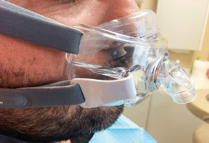
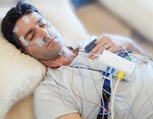
Patient during a polysomnogram (PSG).

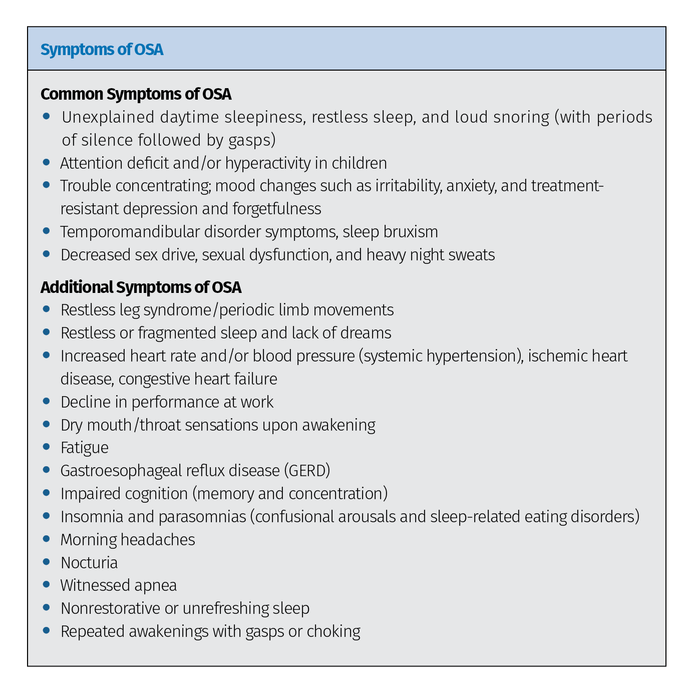
Sleep-related breathing disorders, or SRBDs, are chronic diseases caused by repeated upper airway collapse during sleep. The class includes a range of breathing anomalies, with OSA being the most recognized and most serious of the known SRBDs. OSA is characterized by repetitive episodes of partial or complete obstruction of the patient’s airway during sleep, despite the effort to breathe, and is usually associated with a reduction in blood oxygen saturation. Repetitive reduction of airflow is called hypopnea, and complete cessation of airflow is termed apnea. These episodes of decreased breathing, called apneas (literally meaning “without breath”), typically last 20 to 30 seconds. The level of OSA is described by the Apnea-Hypopnea Index (AHI), which is a count of respiratory events divided by the time of testing. A patient’s AHI is derived using a polysomnogram (PSG), or sleep study, which measures several physiologic aspects to determine the quality of a patient’s sleep. PSGs record changes in brainwaves, eye movements, muscle tone, heart rate, leg movements, and respiration. A sleep physician then reviews the data recorded by the PSG, identifies any anomalies, and makes the appropriate diagnosis.

Prevalence of SRBDs and sleep apnea syndrome, according to age and sex, as reported by the HypnoLaus Study in 2015. (a) A mild SRBD was defined as between 5 and 14 events per hour, while severe SRBD was defined as at least 30 events per hour. SRBD categories differed by age (P < .0001 in men and in women). (b) Mild sleep apnea syndrome was defined as between 5 and 14 events per hour and an Epworth score greater than 10, moderate sleep apnea syndrome was defined as between 15 and 19 events per hour and an Epworth score greater than 10, and severe sleep apnea syndrome was defined as at least 30 events per hour and an Epworth score greater than 10. Categories of sleep apnea syndrome differed by age (P < .0001 in men and P < .001 in women).

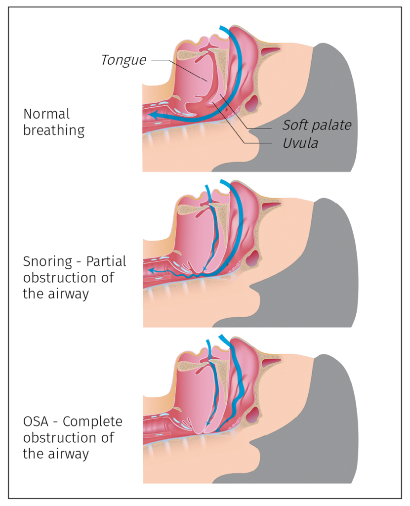
Illustration of the airway during normal breathing, snoring, and OSA.

OSA is largely an anatomical problem. The physical characteristics of the human airway that make complex vocalization possible—our flexible upper airway and suspended mandible—also place us at risk for OSA. During sleep, muscle tone decreases, which can allow inspiratory pressure to collapse into the airway during sleep. The mandible and hyoid bones are suspended, allowing the tongue, soft palate, uvula, and epiglottis to collapse into the airway, creating a complete or partial obstruction superior to the thyroid cartilage and inferior to the hard palate in susceptible individuals. Added risk comes from the fact that, as humans have evolved, the human cranium has increased in size and capacity while the craniofacial respiratory complex has decreased in size. As a result of the smaller oral cavity, the tongue has been progressively displaced posteriorly into the airway, a problem that is exacerbated during sleep. The collapsible nature of the upper airway coupled with a narrow maxilla, small mandible, and displaced tongue has placed humans at great risk for airway obstruction. These anatomical issues can be exacerbated by obesity because as patients become larger, fat deposits in the base of the tongue and the lateral walls of the airway enlarge as well. It is estimated that 70% of OSA sufferers are obese.

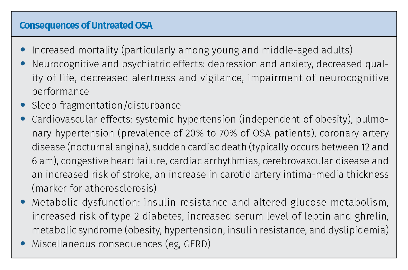
OSA can be deadly. In patients with OSA, hypoxia during apneic episodes results in a sympathetic response and the release of cortisol, initiating the fight or flight response and contributing to sleep disruption and daytime sleepiness. Additionally, the carotid bodies, a small cluster of chemoreceptors and supporting cells located near the bifurcation of the carotid arteries, detect changes in the oxygen/carbon dioxide composition of arterial blood. A decrease in the partial pressure of oxygen and increase in partial pressure of carbon dioxide activates the sympathetic nervous system, which causes the patient’s heart rate to elevate, blood pressure to increase, and respiratory effort to increase. The effect of these sympathetic episodes is an increase in chronic disease and shortened life expectancy in untreated OSA patients.

The majority of individuals with sleep disorders do not present to the physicians for treatment of their sleep issues, but many of these patients will present for routine dental examination and care. A dentist who is properly trained can provide life-saving referrals.

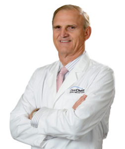
Ken Berley, DDS, JD
Steve Carstensen, DDS, director of dental sleep education for both The Pankey Institute and Spear Education and a close colleague of Dr Berley, chimes in. “I don’t think we’ve reached a point where most dentists are fully aware of the medical consequences of OSA. I believe most understand there is something to snoring and OSA that involves dentists, but the learning curve about medical comorbidities has kept many from putting the information into practice. My dental training includes The Pankey Institute, which emphasizes a comprehensive approach to patient health. While Dr Pankey did not include airway as part of his evaluation, it fits perfectly into a whole-person approach to dental care. Many diagnoses routinely made by dentists have an airway component that must be incorporated into any treatment plan.”

“All dentists must become knowledgeable in the treatment and screening of sleep disorders,” adds Dr Berley, who also uses his extensive law knowledge and experience to advise clinicians on developing their dental sleep medicine practices. “The new ADA statement on the role of dentists in the treatment of SRBDs states that dentists should be screening for OSA, meaning screening for SRBDs will be the standard of care for dentists within a short period of time. From a legal perspective, once the ADA issued the policy statement of the role of dentists in the treatment of SRBDs, plaintiffs’ attorneys will utilize that policy statement as a learned treatise in court to establish a standard of care. Also, I am currently working with several state boards of dentistry to establish their own policy statements that are in line with the ADA. Therefore, within a short period of time, dentists will be legally responsible for conducting a screening during each examination to check for airway insufficiency.”

Adapted from Ramar et al.

“Dentists are in a unique position to change the lives of millions of patients,” Dr Berley continues. “Statistics show that dentists routinely evaluate approximately 50% of the American population each year. It is estimated that 24% of adult men, 9% of adult women, and 10% of children in the US suffer from OSA, and it is estimated that 80% to 90% of all patients who suffer from this devastating disease remain undiagnosed. It is also estimated that each dental office in the United States may have as many as 500 or more patients with undiagnosed OSA currently being treated for dental-related concerns. Therefore, dental professionals have never been presented with a better opportunity to positively impact the health of our patients, but studies have consistently shown that many dentists are unable to recognize systemic and oral risks of OSA. This educational deficiency must be corrected.”

Treating OSA: How Dentists Can Be Involved

The current therapy of choice for OSA is continuous positive air pressure (CPAP) therapy. CPAP works by administering a constant and continuous stream of air under pressure through a mask that is worn over the nose or nose and mouth. The pressure is set to prevent collapse in the oropharynx from negative respiratory pressure and can be customized. When used, CPAP is the most effective means of preventing closure of the airway and thus reducing AHI. But adherence to the therapy is a big problem.

“There are many reasons why people don’t use CPAP,” Dr Carstensen explains. “Each person chooses for themselves how they will think about their diagnosis and the medical risk it poses for them. They have social reasons to pursue therapy, but any medical treatment that requires their cooperation also demands their commitment. If they don’t believe in the value for themselves, overcoming the troublesomeness of the therapy is going to be a problem. The biggest problem in adherence to CPAP therapy isn’t the device—it’s the patient deciding that the benefit does not outweigh the cons. Recent research has found that patients with low levels of certain SRBDs and mild symptoms are far less likely to adhere to CPAP therapy. Reasons people report to me for not using CPAP include the hassle factor of putting the mask on, discomfort from the mask and straps, air flowing into their eyes, complaints by their bed partner, and the inconvenience of traveling with the CPAP machine. Air being pushed into their stomach, drying of their mouth, noise from the machine, and claustrophobia are other frequent issues.”

Each person chooses for themselves how they will think about their diagnosis and the medical risk it poses for them.

Steve Carstensen, DDS
Dr Berley provides specific examples of patients he treated who refused or could not tolerate CPAP therapy: “A 25-year-old woman with an AHI of 47—who once fell asleep at the wheel and hit a bridge abutment going 70 mph down the interstate—refused CPAP therapy because her boyfriend did not like it. This patient was placing her life in jeopardy by refusing CPAP, but acceptance by her boyfriend was more important. Another patient tried faithfully for 10 years to wear his CPAP. He knew how serious OSA is and that, because he was borderline severe, he could suffer serious medical consequences if his OSA was uncontrolled or untreated. However, he suffered significant sinus infections the entire time he used CPAP. Over 10 years of use, he had 3 sinus surgeries.”

Medicare considers a patient to be compliant with CPAP when it is used 4 hours per night at least 5 nights per week. But if a patient sleeps 8 hours per night, 7 nights per week, then a patient using CPAP for 20 out of 56 total hours of sleep in a week—less than half!—is still considered compliant. Additionally, many studies report ranges as low as 25% full compliance over 1 year. SRBDs can be deadly, so a lack of adherence to CPAP therapy is a serious issue, both on an individual basis for each patient and in the broader scheme of how successful sleep medicine can be in treating conditions like OSA.

Enter dentists. CPAP therapy is the medical solution to OSA; oral appliance therapy (OAT) using a mandibular advancement device (MAD) is the dental solution. MADs prevent obstruction of the airway by advancing the mandible, and with it the tongue, thereby expanding the upper airway. Additionally, while it is being worn, the MAD holds the mandible in a stable position throughout the night. Because they are anchored by the teeth for stability, dentists alone are qualified to treat SRBDs using these devices.

Studies have consistently shown that many dentists are unable to recognize systemic and oral risks of OSA. This educational deficiency must be corrected.

Ken Berley, DDS, JD
“Dentists are the only medical professionals qualified to evaluate the patient’s oral health to determine the appropriateness of this treatment and to minimize the potential side effects of OAT,” Dr Berley explains. “In addition to screening for OSA, properly trained dentists can provide a MAD to assist in treatment for patients who are unable to utilize CPAP. OAT is the basis upon which the field of dental sleep medicine was founded, and this field is exploding primarily because the majority of individuals with sleep disorders do not present to their physicians for treatment of their sleep issues. Instead, these patients typically seek medical care for treatment of the comorbid diseases that are directly associated with sleep-disordered breathing—namely hypertension, fatigue, stroke, cardiovascular disease, and diabetes. Sadly, most physicians do not screen for SRBDs, and so the precipitating factors’ contribution to the patient’s chief complaint frequently go undiagnosed. But many of these patients will present for routine dental examination and care, and a dentist who is properly trained to recognize the signs and symptoms of sleep-disordered breathing can potentially provide life-saving referrals and MAD therapy for their sleep-deprived patients. Fortunately, patients with sleep-disordered breathing exhibit identifiable intraoral signs and symptoms as well as physical and social symptoms. With the appropriate training, dentists are well situated to screen and treat many of these patients.”

So how well does OAT with a MAD work when compared with CPAP?

The treatment the patient will use is always more valuable than the one they reject.

Steve Carstensen, DDS
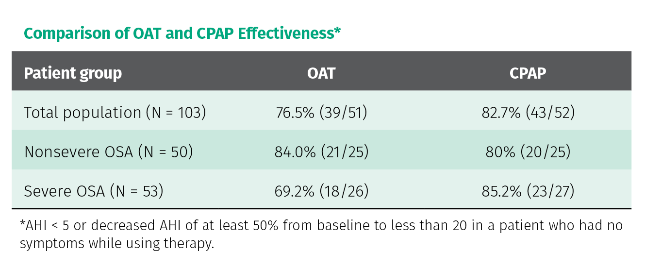
“In an hour-by-hour comparison,” Dr Berley explains, “CPAP is typically more effective at reducing airway closure. Therefore, in most patients, if a CPAP is worn the entire time while sleeping, the patient’s AHI should be less than the same patient who wears a MAD for the same sleep period. In other words, if a patient will wear a CPAP all night, that’s typically the best option to control sleep-disordered breathing. The problem is that most patients cannot or will not tolerate CPAP all night. Patients are typically more compliant during the first part of the night, but by 2:00 am the CPAP is frequently removed. The longest REM sessions occur in the second half of the night, meaning that patients who remove the CPAP midway through the night frequently face the most serious stages of sleep unprotected. Since MADs are typically worn all night, the total amount of protection provided by CPAP and OAT are similar. While it may not be as effective for some patients, if the MAD is worn for 8 hours during sleep versus the CPAP for only 4 hours, OAT results are typically very comparable with CPAP results.

Comparison of OAT and CPAP effectiveness.

“There is more and more research showing that while CPAP therapy is more effective at reducing patient AHI, MAD therapy is more acceptable to more patients,” Dr Carstensen adds. “Physicians and researchers are becoming increasingly aware that the lowest possible AHI is not the only target and that the treatment the patient will use is always more valuable than the one they reject. CPAP therapy is not superior to MAD therapy for many other respiratory details that can be measured, so as the scope of attention widens beyond the most basic measurements, MAD therapy is becoming more accepted for more patients.”

So how did the two patients mentioned above fare when MAD therapy was offered instead of CPAP? The young woman’s AHI was reduced to 1.2, and the MAD was accepted by her boyfriend. The other patient’s AHI was reduced to 0, and his sinus infections completely stopped.

The longest REM sessions occur in the second half of the night, meaning that patients who remove the CPAP midway through the night frequently face the most serious stages of sleep unprotected.

Ken Berley, DDS, JD
“Research shows that some people are more easily aroused from any level of sleep,” Dr Carstensen explains, “so an external device that provides constant sensory signals to the person has the potential to be more of an irritant to sleep than a facilitator of it. Once the patient gets accommodated to the MAD, there are no new sensory signals coming from it to interfere with normal sleep patterns. I have a patient, Susan, who came to me after reading about OAT online. She sought diagnosis 3 years previously, mostly because of unrefreshing sleep and poor work performance. She was diagnosed with mild sleep apnea, with a significant portion of those airway events being hypopnea. She was, nevertheless, given a CPAP. I don’t have the complete details of that course of CPAP therapy, but she said she used two different CPAP machines, more than six masks, and years of trying before abandoning the CPAP altogether. She said her sleep physician did not suggest any alternative courses of treatment, given that her apnea was ‘mild, anyway.’ Susan was very frustrated because that left her in basically the same condition she was in prior to therapy, with 3 years and tons of effort down the drain.

“With the medical records from the sleep physician came a prescription for an oral appliance,” he continues. “Susan was happy to move forward and was soon accommodated to the device, which she immediately felt was better than her CPAP. We did send her home with monitors to refine her jaw position, but we were able to achieve a normal AHI and eliminated all flow limitations within 8 weeks of meeting her. Her subjective symptoms completely resolved once we had her jaw postured correctly. She was absolutely thrilled we were able to reach this positive outcome so quickly, contrasting it with her 3-year trial of CPAP therapy, and she was a little annoyed that her sleep physician didn’t suggest this in the first place.”

The treatment plan also doesn’t have to be an either/or decision with clinicians choosing between the advantages of MAD therapy or the advantages of CPAP therapy, weighed against the disadvantages of both.

A patient is fitted for a MAD that will be used in combination with his CPAP.

“For patients who are religiously trying to comply with CPAP but are unable to wear their device all night,” Dr Berley explains, “we can provide a MAD to be worn in conjunction with CPAP. We call this combination therapy. In this situation, the oral appliance is placed in the mouth, then the CPAP is positioned. The MAD helps open the patient’s airway so that the pressure setting on the CPAP can be reduced below what it would need to be set at without the MAD, thereby making the CPAP easier to wear. This provides the patient with the best possible outcome. The oral appliance is typically left in place even if the CPAP is removed during the night, meaning the patient still has a level of protection. Additionally, the MAD can be used in times of power outages and off-the-grid situations.”

Dr Carstensen gives an example of this treatment plan in practice: “April was referred from a physician after a sleep lab experience. The test showed severe OSA, so, as was the order, the sleep technologist woke April to place a CPAP and see if that could help her. The mask lasted fewer than 30 seconds, and April left the room immediately. She said, ‘I’d rather die in my sleep than use that mask.’ We made her a well-fitted, well-tolerated oral appliance that, sadly, was not effective enough at controlling her OSA. I needed her to be able to use a CPAP, probably in combination with the oral device, so I referred her to a sleep-aware psychiatrist who helped April get past some PTSD issues that were triggering her CPAP aversion. Afterward, April became a new person. She felt great, had a better frame of mind, and was happily using her CPAP device most nights and her oral appliance on weekends and for traveling. She could not have achieved this excellent health outcome without a team of involved providers willing to find the right solution for her rather than focusing only on their own areas of expertise. It was rewarding knowing that I facilitated this happy outcome—an experience that was uncommon in most restorative dentistry cases.”

(a and b) Herbst appliance. (c) EMA appliance. (d) dreamTAP appliance. (e) ProSomnus [IA] appliance. (f) Dorsal fin appliance. (g) ProSomnus [CA] appliance. (h) Moses appliance.

Working with Our Partners in Medicine

So how do you dive in? Dental sleep medicine is a growing field of dentistry, but the amount of ‘new tricks’ to learn has kept many dentists from stepping out of their comfort zone. In addition to the learning curve that applies to implementing any new form of treatment, dental sleep medicine is also uniquely challenging in that it involves careful cooperation with a board-certified sleep physician. Because OSA is considered a medical disease, dentists cannot legally diagnose it. The PSG testing and diagnosis instead must come from a sleep physician; after diagnosis, treatment either comes from the sleep physician by way of CPAP therapy or, with physician referral, treatment with OAT from a trained dentist. It’s a complicated dance between the fields of medicine and dentistry, but dentists shouldn’t feel overwhelmed. The path is possible, and, in fact, the junctions with medicine actually offer some advantages to dentists.

“Because OSA is a medical condition,” Dr Berley explains, “OAT is filed on the patient’s medical insurance. There are no dental codes available. This presents a major opportunity to provide OAT and not utilize your patient’s dental insurance benefits. The treatment of OSA with MADs can also provide an additional income stream for offices wishing to expand their services. Many practitioners who provide OAT are routinely billing medical insurance for payment. All diagnosis and treatment planning for SRBDs/OSA is provided by a sleep physician, who controls the treatment and determines when OAT is appropriate for a patient, then refers that patient to a dentist for OAT.”

Dentists trained in dental sleep medicine can be a vital member of a multidisciplinary team of health care professionals working together to control this deadly disease.

Ken Berley, DDS, JD
After referral for treatment by a sleep physician, the dentist then takes on the role of durable medical equipment provider, filling the physician’s prescription with an oral appliance. But, while that title may minimize the dentist’s role in treatment, the reality is that OAT requires a rigorous amount of clinical and treatment-planning expertise from the cooperating dentist, as well as extensive follow-up care to manage treatment and watch for any dental complications. Cooperation with sleep physicians becomes a two-way street: As dentistry accepts the mantle of SRBDs/OSA screening, it becomes all the more important to have trusting relationships with sleep physicians to whom they can send patients. And, in return, dentists may also receive patients who have come to the sleep physician on their own or through other specialists treating the myriad comorbid conditions associated with SRBDs. As dental sleep medicine expands, it strengthens dentistry’s role in a collaborative effort to address a serious, widespread disease.

“SRBDs directly or indirectly affect many biologic systems within the human body,” Dr Berley explains. “Therefore, numerous medical disciplines are involved in the treatment of OSA comorbid conditions. This fact has allowed dentists to develop an extensive physician-based referral practice. Dentists trained in dental sleep medicine can be a vital member of a multidisciplinary team of health care professionals working together to control this deadly disease.”

There’s a lot to learn before you break into dental sleep medicine. To both help and encourage fellow dentists to get involved, Drs Berley and Carstensen have coauthored The Clinician’s Handbook of Dental Sleep Medicine, which effectively serves as a comprehensive how-to manual for starting a dental sleep medicine practice. All of the information clinicians need is covered: the current scientific knowledge of SRBDs, the clinical information needed to provide and deliver OAT, guidance for filing OAT on a patient’s medical insurance or Medicare, and the legal information necessary for navigating a complicated system at the boundary of dentistry and medicine.

“What I want is for every dentist to be aware of SRBDs, look for them in their practice, and facilitate a solution for them,” Dr Carstensen states. “I grew up with my father being an epic snorer, thinking nothing about it in connection with health. With my knowledge now of airway, snoring, sleep apnea, and the effects of these problems, I don’t want dentists to think their family members and patients are simply snorers. My father has severe sleep apnea; after I finally got him to a sleep doctor, he began using a CPAP device and enjoyed his most productive years at work in the few remaining before retirement. After retiring, he was able to drive safely around North America in his RV for the next 17 years. If our book helps clinicians decide that they can treat patients like my father, they can share the rewards that come from truly making a difference in these patients’ lives. This positive work will generate rewards in their own lives, as well.”

“While I love to lecture and show the amazing clinical results of OAT, the real joy is hearing the stories of how our therapy changes the lives of our patients,” Dr Berley adds. “My favorite story is of one of the first patients I treated. He was 32 years old when he was first diagnosed. He was an athletic young man whom you never would have dreamed was a severe apneic, but he had a very small oral cavity secondary to four orthodontic premolar extractions. His AHI was 54, and he snored like a freight train. While the snoring was inconvenient, it never caused him many problems—until he got engaged. Suddenly, the snoring was the most important thing in his life. Shortly after he was married, he was in trouble. He was tested for OSA and placed on CPAP. He tried desperately to comply with CPAP treatment, but to no avail. Within 6 months of being married, he was relegated to the guest bedroom, and the couple was in marriage counseling. The wife could not understand how difficult it can be for some patients to comply with CPAP. After OAT, his AHI was reduced to 0.4, but—more importantly—his snoring completely stopped. The couple now has three children, and the wife will personally call me to let me know when I need to adjust her husband’s appliance.

“My journey of study into OSA culminated in two things,” Dr Berley concludes. “As a patient, my journey led me to the Herbst appliance that I wear each night. As a clinician, my journey led me to The Clinician’s Handbook of Dental Sleep Medicine. I hope my experience gives readers a small glimpse into the satisfaction of providing OAT for the treatment of SRBDs, and I hope our book gives clinicians the courage to start their own journeys. Had it not been for the introduction of dental sleep medicine into my practice protocol, I would have retired several years ago. Treating OSA has made dentistry fun again. If you’re looking for something to reinvigorate your passion for dentistry, take a good look at dental sleep medicine—you will not be disappointed. And if there is anything I can do to assist you, please feel free to contact me.”
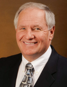

Ken Berley, DDS, JD, is a dentist and lawyer who, for the past 10 years, has focused his dental practice on the treatment of sleep-disordered breathing and temporomandibular disorders. He is also the president of a consulting firm offering in-office training on dental sleep medicine (DSM) and consent forms and documents to assist the DSM practice. Dr Berley is a Diplomate of the American Board of Dental Sleep Medicine and regularly lectures in the areas of risk management and the development of a successful DSM practice. He has been published in Sleep Review magazine and other journals associated with sleep-disordered breathing, and he provides consulting services for various insurance companies and actively defends and advises dentists who are facing legal challenges.
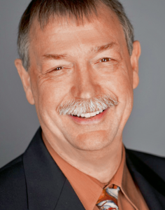
 

Steve Carstensen, DDS, started treating sleep problems in 1998 and maintains a private practice in Bellevue, Washington. He has completed a Mini-Residency in Sleep at the University of California, Los Angeles, and is a Diplomate of the American Board of Dental Sleep Medicine. He lectures internationally, directs sleep education at the Pankey Institute, and is a guest lecturer at Spear Education, University of the Pacific, and Louisiana State University Dental Schools. Since 2014, he has been editor-in-chief of Dental Sleep Practice Magazine.
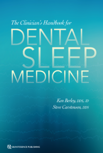
 

The Clinician’s Handbook for Dental Sleep Medicine

Ken Berley and Steve Carstensen

It has been estimated that 20 million Americans suffer from moderate to severe OSA, and at least one patient in five has mild OSA. The primary treatment prescribed by sleep physicians is CPAP, but patient compliance with this therapy is unacceptably low, between 25% and 50%. There is a significant opportunity for dentists to provide a viable alternative therapy—oral appliance therapy (OAT). OAT results in much better adherence to therapy than CPAP, and while OAT is not as efficacious as CPAP, this increased compliance results in comparable therapeutic results. Currently, a board-certified sleep physician is the only medical professional qualified to diagnose OSA and other sleep-related breathing disorders (SRBDs), so dentists must coordinate with a sleep physician to provide OAT. This book is the how-to guide, a gateway to a successful dental sleep medicine practice. Written by two experts in the field, it clearly delineates the dentist’s role in the treatment of SRBDs and gives practical advice for how to incorporate dental sleep medicine into an existing dental practice, not to mention how to work with sleep physicians to best support patient care. In addition to step-by-step instructions for examination, appliance selection, and follow-up care, complications of OAT, legal issues, and medical insurance and Medicare considerations are included to fully prepare the dentist for the journey into dental sleep medicine.

240 pp (softcover); 60 illus; ©2019; ISBN 978-0-86715-813-7 (B8137); Now available! $72


This article was written by Caitlin Davis, Quintessence Publishing.

©2019 BY QUINTESSENCE PUBLISHING CO, INC. PRINTING OF THIS DOCUMENT IS RESTRICTED TO PERSONAL USE ONLY. NO PART MAY BE REPRODUCED OR TRANSMITTED IN ANY FORM WITHOUT WRITTEN PERMISSION FROM THE PUBLISHER.

Posted in Books, Feature, Multidisciplinary, Sleep Dentistry | Tagged , , , , , , , , , , | 5 Comments

ISPRD 2019 Spotlight: Dental Photography with Miguel A. Ortiz, DMD

Reading time: 3 minutes

Dr Miguel A. Ortiz will be presenting an all-day hands-on workshop on Thursday, June 6, 2019, during the presymposium portion of The 13th International Symposium on Periodontics & Restorative Dentistry.

My goal in teaching this workshop is to develop attendees’ general and clinical photography skills by teaching them the fundamental concepts of photography. Every photographer must have a strong grasp of these concepts in order to optimize their work. There is no simple formula of settings that will always work because any slight change in physical space, lighting, angle, or distance can drastically change the photographic outcome. However, once you understand the physical and mechanical aspects behind photography, you can approach each situation with the knowledge of how to adjust the camera’s settings to achieve your goals. You learn the language of lighting and can manipulate it the way great musicians manipulate their instruments to draw out an infinite spectrum of texture, tone, and range. The best way to learn these concepts is through a combination of clear, concise didactic instruction and plenty of hands-on practice guided by photography specialists.

Photography is like driving a car. I can tell you what all the buttons are and what they do, but until you get behind the wheel yourself, you won’t know how to drive. So you practice. You sit behind the wheel with an experienced instructor beside you to guide you as you take those first slow loops around the block. Reading and hearing someone talk about driving may give you knowledge, but it doesn’t provide the experience of adjusting to different situations. Like a new driver getting onto the freeway for the first time, you need that guided practice to adjust your speed and merge into oncoming traffic. My workshop offers that same guided practice for dental photography. You learn to gauge what each photographic setting needs with your own eyes, your own hands, and your own experience. You take the concepts you’ve learned and practice applying them with live models to explore positioning and how to verbally guide the subject. You will work with multiple lighting stations, equipment, and setups, giving you the chance to evaluate a range of equipment to see what you like and what works best for your needs.

Anyone and everyone in the dental field will benefit from this course. All members of the dental team, including assistants, hygienists, and office staff, are an integral part of maintaining accurate and consistent photographic protocols. Dental students and residents especially will benefit from the skills learned in this course by using high-quality photography of their work in developing, documenting, collaborating, and assessing treatment plans and patient care.

The value of this workshop is in building a strong foundational understanding of photography that can be applied in life and in work. Attendees take home a simple protocol for intra- and extraoral photographs and in-depth knowledge of equipment and lighting setups for clinic space optimization. They will come out of the workshop with confidence in their skills as a photographer gained through the extensive hands-on experience.

Miguel A. Ortiz, DMD
Dental Photography Course
An All-Day Hands-On Workshop at ISPRD 2019
Register today at www.bostonperiomeeting.com

Posted in Announcement, Multidisciplinary | Tagged , | Leave a comment

Quintessence Roundup: February

Reading time: 12 minutes

New Titles in Books


The Clinician’s Handbook for Dental Sleep Medicine

Ken Berley and Steve Carstensen

It has been estimated that 20 million Americans suffer from moderate to severe OSA, and at least one patient in five has mild OSA. The primary treatment prescribed by sleep physicians is CPAP, but patient compliance with this therapy is unacceptably low, between 25% and 50%. There is a significant opportunity for dentists to provide a viable alternative therapy—oral appliance therapy (OAT). OAT results in much better adherence to therapy than CPAP, and while OAT is not as efficacious as CPAP, this increased compliance results in comparable therapeutic results. Currently, a board-certified sleep physician is the only medical professional qualified to diagnose OSA and other sleep-related breathing disorders (SRBDs), so dentists must coordinate with a sleep physician to provide OAT. This book is the how-to guide, a gateway to a successful dental sleep medicine practice. Written by two experts in the field, it clearly delineates the dentist’s role in the treatment of SRBDs and gives practical advice for how to incorporate dental sleep medicine into an existing dental practice, not to mention how to work with sleep physicians to best support patient care. In addition to step-by-step instructions for examination, appliance selection, and follow-up care, complications of OAT, legal issues, and medical insurance and Medicare considerations are included to fully prepare the dentist for the journey into dental sleep medicine.

240 pp (softcover); 60 illus; ©2019; ISBN 978-0-86715-813-7 (B8137); $72 Special preorder price! $58
Available March 2019

Read more about The Clinician’s Handbook for Dental Sleep Medicine here!

Obstructive Sleep Apnea: A Deadly Disease with a Dental Solution


Surgical Management of Maxillofacial Fractures

Yoh Sawatari

The facial skeleton is comprised of vertical and horizontal buttresses and the intersections they create; maxillofacial fractures occur when these buttresses sustain more force than they can withstand. The objective when managing these fractures is to reverse the damage that these buttresses sustained and restore appropriate facial dimensions. Not all fractures propagate in the same pattern, so surgeons must compartmentalize the face and define the character of the individual bones. This book approaches the face one bone at a time, outlining how to evaluate each type of fracture, the indications for surgery, the surgical management, and any complications. Specific protocols for clinical, radiographic, and CT assessment are included, as well as step-by-step approaches for surgical access and internal reduction and fixation. Isolated fractures are rare with maxillofacial trauma, and the author discusses how to sequence treatment for concomitant fractures to ensure the most successful outcome. This book is a must-have for any surgeon managing maxillofacial fractures.

256 pp; 254 illus; ©2019; ISBN 978-0-86715-794-9 (B7949); Now available! $178


Dental Anatomy and Morphology

Hilton Riquieri

This beautiful atlas conveys not only the practical knowledge of dental anatomy but also the art of sculpting it in wax. The ideal anatomy of each dental structure is described in detail and the waxing techniques are beautifully illustrated step by step for visual reference. The author demonstrates that for every morphologic feature there is an explanation in nature, assigning significance to every minute feature of dental morphology. Organized by tooth and arch, this book views morphology through a clinical lens and repeatedly draws connections between anatomical features and clinical concepts. The fundamental knowledge presented in this text is essential for improving waxing and sculpting techniques and will be useful for students and specialists alike.

332 pp; 1,507 illus; ©2019; ISBN 978-0-86715-770-3 (B7703); Now available! $172


Quintessence of Dental Technology 2019: Volume 42

Edited by Sillas Duarte, Jr

QDT 2019 presents a potpourri of original articles highlighting new techniques and novel approaches for creating beautiful smiles—both in form and function. Featured articles include an innovative procedure for predictably matching a veneer to an implant crown, a program for producing a personalized smile based on its visual identity, and the Plane System for virtual functional and esthetic analysis, diagnosis, and CAD/CAM fabrication. These are but a few of the pearls found in this year’s beautifully produced annual resource for the dental technician and restorative clinician.

232 pages; 1,000+ illus; ©2019; ISBN 978-0-86715-816-8 (JQ630); Now available! $156


Técnica de Alineadores Transparentes

Sandra Tai

Los alineadores transparentes son el futuro de la ortodoncia, pero la ortodoncia digital evoluciona tan rápidamente que es difícil estar al día. Este libro trata sobre el uso de alineadores transparentes desde una perspectiva de diagnóstico y planificación del tratamiento; también, discute cómo se aplican principios de biomecánica y anclaje en el uso de estas técnicas. Cada capítulo explica cómo usar alineadores transparentes para tratar una oclusión no óptima y enseña a los expertos a diseñar un tratamiento adecuado usando el software disponible, así como la manera de hacer el tratamiento clínico y terminarlo. Este manual clínico preparará a los ortodoncistas y estudiantes dentales a superar las expectativas del paciente con los aparatos ortodónticos más estéticos disponibles hoy en día.

308 pp; 1,344 ilus; ©2019; ISBN 978-0-86715-789-5 (E7895); Now available! $118

Read more about Técnica de Alineadores Transparentes here!

Clear Aligner Technique: Bringing Orthodontic Treatment into the Digital Era


The Oral-Systemic Health Connection: A Guide to Patient Care, Second Edition

Edited by Michael Glick

As the oral-systemic health connection continues to be the focus of an overwhelming amount of scientific literature, it is important for dentists to stay informed in order to be a reliable source of information for their patients regarding both oral and overall health. This second edition of The Oral-Systemic Health Connection: A Guide to Patient Care explores the connections between oral infections and systemic diseases/conditions, incorporating feedback from scientists, practitioners, and policymakers. In addition to updated chapters about cardiovascular disease, diabetes, inflammation, and adverse pregnancy outcomes, authors have contributed new chapters about antibiotic prophylaxis, the genomic connection, common risk factors, and the economic impact of this connection. Because interpreting this research can be challenging, new chapters about causal frameworks and biostatistical assessment have been added, and specific clinical considerations for providing dental care to patients with certain conditions have been included in selected chapters. Besides providing readers with up-to-date information on the complex connection between oral and general health, this book prepares oral health care professionals to critically read and evaluate new research to ultimately benefit their patients’ overall health.

384 pp (softcover); 85 illus; ©2019; ISBN 978-0-86715-788-8 (B7888); Now available! $48


Next-Generation Biomaterials for Bone & Periodontal Regeneration

Edited by Richard J. Miron and Yufeng Zhang

New and innovative biomaterials are being discovered or created in laboratories at an unprecedented rate, but many of them remain entirely foreign to practicing clinicians. This book addresses this gap in knowledge by summarizing some of the groundbreaking research performed to date on this topic and providing case examples of these biomaterials at work. The book begins with a review of the biologic background and applications of bone grafting materials utilized in dentistry. The principles of guided tissue and bone regeneration are covered in detail, including many recent advancements in barrier membrane technologies as well as use of platelet-rich fibrin and various growth factors, and many next-generation materials that will optimize future bone and periodontal regeneration are presented. The final chapter is designed to help clinicians select appropriate biomaterials for each specific regenerative protocol. Much like one implant size and shape cannot be utilized for every indication in implant dentistry, one bone grafting material, barrier membrane, or growth factor cannot maximize regenerative outcomes in all clinical situations. This textbook teaches clinicians how to utilize biomaterials in an appropriate, predictable, and evidence-based manner.

384 pp; 960 illus; ©2019; ISBN 978-0-86715-796-3 (B7963); Now available! $218

Read more about Next-Generation Biomaterials for Bone & Periodontal Regeneration here!

Next-Generation Biomaterials: What They Are, Why You Need to Know About Them, and Which Ones Should Intrigue You


The Sinus Bone Graft, Third Edition

Edited by Ole T. Jensen

As research proceeds on treatment of the resorbed posterior maxilla, new techniques and innovations continue to be adopted to solve this clinical problem. While the previous edition of this book provided detailed information on the types of grafting materials and procedures available at the time, this completely revised version looks to the future with new strategies for treatment, some of which avoid grafting altogether. This book not only reviews the time-tested lateral window approach for sinus elevation and grafting but also describes a variety of techniques to approach the sinus transcrestally with or without grafting material. One section of the book is devoted entirely to the different types of implants and implant placement techniques available, many of which are designed specifically to avoid sinus elevation. In addition to clinical case studies and descriptions of how to perform specific surgical procedures, this book includes discussions on the science of bone formation and how continued research brings us closer every day to the ultimate goal of using tissue engineering to completely regenerate new teeth.

288 pp; 948 illus; ©2019; ISBN 978-0-86715-791-8 (B7918); Now available! $168

Read more about The Sinus Bone Graft, Third Edition here!

Introducing The Sinus Bone Graft, Third Edition, edited by Ole T. Jensen


The Ortho-Perio Patient: Clinical Evidence & Therapeutic Guidelines

Edited by Theodore Eliades and Christos Katsaros

Although most orthodontic curricula provide courses on interdisciplinary orthodontic-periodontic treatment, there are still surprisingly few resources on the topic. Written by leading scholars in the field, this book provides a broad analysis of the topic from both the periodontal and orthodontic perspectives. The authors systematically analyze the scientific and clinical interactions of these specialties by reviewing all the available evidence and using case studies to demonstrate principles discussed in theory. The result is a text that outlines the treatment fundamentals and shows how to improve the therapeutic outcomes involving orthodontic-periodontic interventions.

224 pp; 346 illus; ©2019; ISBN 978-0-86715-679-9 (B6799); Now available! $128


3D Printing in Dentistry 2019/2020

Irfan Ahmad and Fahad Al-Harbi

The invention of 3D printing technology and its microprecision products are revolutionizing the way dentistry is practiced. However, as with any new technology, many clinicians are uncertain of where to start. The purpose of this book is to arm clinicians with what they need to know to incorporate 3D printing into daily practice. After a brief discussion of the evolution of 3D printing and the scientific credence behind it, the first section examines the fundamental concepts and the latest trends in digital dentistry, including intra-oral scanning, 3D printers, 3D materials, and CAD/CAM processes. The second half of the book presents clinical case studies that apply 3D printing to treat surgical, orthodontic, prosthodontic, restorative, and esthetic cases. These cases examine the benefits and limitations of 3D printing technology in density. This text is a must-have for dentists wanting to understand the future of dentistry.

256 pp; 390 illus; ©2019; ISBN 978-1-78698-026-7 (BL083); Now available! $138


Restauración con fluidas

Douglas A. Terry

Este libro contiene las distintas aplicaciones de los compuestos de resina fluidos de nueva generación y las presenta paso a paso. Los primeros capítulos tratan sobre su evolución y la ciencia que respalda el concepto de su diseño adhesivo y la técnica compuesta de resina inyectable. Los capítulos siguientes presentan casos en los que se recurre a su uso clínico, como restauraciones anteriores y posteriores, coronas pediátricas, adhesiones de restauraciones indirectas, desarrollo del sitio de póntico ovoide, eliminación de la sensibilidad en el cervical del diente, sellado inmediato de la dentina, reparación de dentadura fracturada, entre otros. Cada presentación de caso incluye los varios diseños de adhesivos preparativos, técnicas reconstituyentes, protocolos adhesivos y los procedimientos finales correspondientes. Con el uso de este material ampliará las opciones de tratamiento dental, la precisión y la predictibilidad, reduciendo el tiempo del paciente en su consultorio.

292 pp; 914 illus; ©2019; 978-0-86715-775-8 (E7758); Now available! $118

Read more about Restauración con fluidas here!

Flowable Composite Resins: Esthetics at Your Fingertips


Kratochvil’s Fundamentals of Removable Partial Dentures

Ting-Ling Chang, Daniela Orellana, and John Beumer III

In the 1960s, Professor F. J. Kratochvil recognized the importance of biomechanics in removable partial denture (RPD) design and used these principles to develop a new design philosophy. This “RPI system”—a clasp assembly consisting of a rest, a proximal plate, and an I-bar retainer—changed how clinicians approach partial denture design and is now used throughout the world. This textbook provides an overview of Kratochvil’s design philosophy and the basic principles of biomechanics it is based upon. Topics include components of RPDs and their functions, design sequences for maxillary and mandibular RPDs, and techniques for surveying and determining the most advantageous treatment position. A chapter dedicated to digital design and manufacturing of RPD frameworks highlights new technology in this emerging field. Additional topics include optimizing esthetic outcomes through attachments and rotational path RPDs as well as applying the RPI system to patients with maxillofacial defects. The authors provide illustrations of clinical cases throughout the book as well as an illustrated glossary of prosthodontic terminology. This textbook will prepare students and general practitioners to design and fabricate a biomechanically sound RPD framework for just about any dental configuration they encounter.

240 pp; 748 illus; ©2019; ISBN 978-0-86715-790-1 (B7901); Now available! $108

Read more about Kratochvil’s Fundamentals of Removable Partial Dentures here!

Kratochvil’s RPD Design Principles Five Decades Later: How Today’s Prosthodontists Continue to Build Upon His Legacy


Current Issues in Journals


prd_banner

Featured article: Evaluation of the Combination of Strip Gingival Grafts and a Xenogeneic Collagen Matrix for the Treatment of Severe Mucogingival Defects: A Human Histologic Study
Istvan A. Urban, Katalin Nagy, Sabine Werner, and Michael Meyer

Randomized Controlled Clinical Trial of All-Ceramic Single-Tooth Implant Reconstructions Using Modified Zirconia Abutments: Results at 5 Years After Loading
Andrea Laass, Irena Sailer, Jürg Hüsler, Christoph H. F. Hämmerle, and Daniel S. Thoma

The CAD/CAM Compound Prosthesis: Digital Workflow for Fabricating Cement-Retained Zirconia Prosthesis Over Screw-Retained Milled Titanium Bars
Meisam Faeghinejad, Periklis Proussaefs, Abdulaziz AlHelal, and Jaime Lozada

Assessment of Hypodontia Treatment of Maxillary Lateral Incisors in Adult Patients After 9 Years of Follow-up: A Retrospective Study
Barbara Rafałowicz and Leopold Wagner

Retrospective 9-Year Clinical Outcome Report on Adhesive Post-endodontic Treatment of Anterior Teeth Using Prefabricated Fiber Posts
Daniel Cerny, Steven Eckert, and Radek Mounajjed

Implant Inclination and Cantilever Length Are Not Associated with Bone Loss in Fixed Complete Dentures: A Prospective Study
Bernardo A. Camargo, Luís G. R. Drummond, Ahmet Ozkomur, Eduardo A. Villarinho, Maria Ivete B. Rockenbach, Eduardo R. Teixeira, and Rosemary S. A. Shinkai

jomi_banner

Thematic abstract review: Factors Associated with Oral Implant Failures
Emad W. Estafanous

In Vitro Study of Bond Strength Between Abutments with Different Degrees of Convergence and Crowns by Pre-Bonding Method
Liguo Zhao, Paul Weigl, Yanyun Wu, and Yuanzhi Xu

Effect of Different Bar Designs on Axial and Nonaxial Retention Forces of Implant-Retained Maxillary Overdentures: An In Vitro Study
Moustafa Abdou ELsyad, Radwa M. K. Emera, and Tarek Mohy Ashmawy

Nortriptyline Compared to Amitriptyline for the Treatment of Persistent Masticatory Myofascial Pain
Yaron Haviv, Avraham Zini, Yair Sharav, Galit Almoznino, and Rafael Benoliel

Tooth Clenching Until Exhaustion Evokes Exercise-Induced Hypoalgesia in Healthy Persons and in Patients with Temporomandibular Disorders
Sarah Vaderlind Lanefelt, Mauricio Mélo-Gómez, Mariam Chizari, Mirna Krsek, Nikolaos Christidis, Eva Kosek, and Malin Ernberg

A Prediction Model for Types of Treatment Indicated for Patients with Temporomandibular Disorders
Naichuan Su, Corine M. Visscher, Arjen J. van Wijk, Frank Lobbezoo, and Geert J.M.G van der Heijden

Finishing with clear aligner appliances: A systematic review
Beatriz Solano Mendoza, Galder Hernando Martín, and Carolina Caleza Jiménez

Comparison of tooth movement with aligners with and without acceleration devices. Part 2: Oral health-related quality of life and pain in patients with acceleration devices
Xianju Xie, Hongyang Yin, Werner Schupp, Julia Haubrich, Hanna Gerwing, and Yuxing Bai

Molar distalization 2 by 2
Patrice Bergeyron


Dental Meetings Quintessence Will Attend in February


Yankee Dental Congress: Booth #1003
hosted by the Massachusetts Dental Society January 31–February 2 in Boston, Massachusetts

ICOI Winter Implant Symposium: Booth #101
hosted by the International College of Oral Implantologists February 14–16 in Phoenix, Arizona

CDS 2019 Midwinter Meeting: Booth #3311
hosted by the Chicago Dental Society February 21–23 in Chicago, Illinois

AAFP 68th Annual Scientific Session
hosted by the American Academy of Fixed Prosthodontics February 22–23 in Chicago, Illinois

LMT LAB DAY Chicago
hosted by LMT Communications February 21–23 in Chicago, Illinois


Upcoming Quintessence Events


Posted in Books, Journal of Oral & Facial Pain and Headache, Journals, Promotions, Roundup, The International Journal of Oral & Maxillofacial Implants, The International Journal of Periodontics & Restorative Dentistry, The International Journal of Prosthodontics, What's New | Leave a comment

Next-Generation Biomaterials: What They Are, Why You Need to Know About Them, and Which Ones Should Intrigue You

Reading time: 22 minutes

Biomaterials are a hot topic in modern dentistry. Open any textbook on implant or periodontal surgery, and, in addition to the in-depth descriptions of the surgical protocols, you are bound to find the author’s biomaterial recommendations for each procedure. Gather 20 clinicians in a room and you will likely hear 20 different ways to execute a single procedure. Knowing one way to do something is good—knowing multiple ways to do something is better because it increases versatility and enables the clinician to respond to different clinical indications. That’s the reasoning behind Drs Richard J. Miron and Yufeng Zhang’s new textbook Next-Generation Biomaterials for Bone & Periodontal Regeneration, which takes the opposite approach from other textbooks in that, rather than starting with the clinical indication or surgical procedure and working toward the biomaterial recommendations, they start instead with each biomaterial and work outward from there.

“There are so many textbooks that are on implants or bone grafting or soft tissue management,” says Richard J. Miron, DDS, BMSc, MSc, PhD, Dr med dent, “but there hasn’t really been a textbook that was dedicated to bone and periodontal biomaterials and how and when to use them effectively. When it comes to biomaterials, one thing that is difficult for the average clinician to figure out is that every single company selling biomaterials will tell you their biomaterial can be utilized for every clinical procedure. They promote the perfect all-purpose material, but the reality is that it’s likely not the case. And in textbooks and courses, the author or presenter will provide their clinical preference, but never in the great context of all the available options for that procedure.”

What Drs Miron and Zhang have done differently is break down the biologic background of each biomaterial chapter by chapter. The result is a text that provides the detailed information about each unique biomaterial that will best equip the clinician to use that biomaterial most effectively in any clinical situation for which it is indicated. Again, knowing the steps to do something is good, but knowing the reasoning behind each step is better. If you don’t know how a biomaterial works, you won’t be able to alter how you use it in different clinical indications to achieve the best results.

“One thing that happens,” Dr Miron explains, “is that when people go to dental school, they learn how to do something and get comfortable doing each procedure a certain way and continue doing it that way for the remainder of their careers, which is fine. You always want to perform surgery in an effective way with what works well in your hands and based on your educational and clinical experiences. But because the field of dentistry has been progressing so rapidly, especially in recent years, what happens is what we learned 10 years ago becomes outdated, new tools become available, and new biomaterials facilitate and improve our field, yet it remains difficult to learn all this new knowledge effectively. Furthermore, marketing from companies is at an all-time high, often not providing the best available data or advice to the clinician. We have to close this gap somehow, and the best way is to teach experienced clinicians using evidence-based biomaterials and provide the information they need to make their own rational decisions.”

In their book, Drs Miron and Zhang essentially provide all of the information necessary to solve for one equation: For a given procedure, which grafting material, barrier membrane, and growth factor should you consider to achieve optimal results?

“We have literally gone chapter by chapter on each biomaterial,” Dr Miron explains, “and in each chapter we provide background information that delivers the biologic basis for each biomaterial and how it effectively functions and integrates into the human body. By going through the biologic behavior of each biomaterial, the reader is then taught where and why to use it. We’re not saying ‘This is the clinical situation, so which materials should we use?’ We’re saying ‘This is the material, so let’s explain to you how it works, what regenerative properties it has compared to others, and then determine where we should use it.’ Some of these biomaterials have overlapping indications. Because of that, at the very end of the book we got together with all of the clinicians who helped us write the book and developed a chapter that provides biomaterial recommendations for each clinical indication. On top of that, because more and more clinicians are practicing holistic dentistry in a trend that continues to increase, we have also included alternative options using a holistic approach for every procedure. It’s a different approach with a different perspective that will change how you visualize these biomaterials.”

So if a book like this has never been written before, what made it possible for Drs Miron and Zhang? The answer lies in the unique educational and professional backgrounds of each editor.

“One of the advantages we had in writing this book is that Yufeng Zhang and I are both molecular and cell biologists and dentists,” Dr Miron states, “so we approach each biomaterial from the perspective of both a scientist and a clinician. We both do a lot of preclinical testing on biomaterials, so we have access to all of these different materials in various stages of testing. Since we’ve utilized these materials both preclinically and clinically, it was a good opportunity for us to provide real concrete scientific evidence of each biomaterial and to give our recommendations for their uses. I do not believe any research group anywhere in the world has had such an opportunity to test so many different materials, and we felt it was now necessary to translate what we’ve learned over the past 10 years into an easy-to-read textbook. Our goal was not to simply give recommendations. Our goal was that the dentist better understand each biomaterial to allow them to make better clinical choices for their use as opposed to simply relying on sales reps and vendors to provide their educational information.”

While the number of biomaterials currently available may already seem overwhelming to the clinician, and new ones seem to come to market every other day, this is only the tip of the iceberg. Behind every newly available biomaterial are scores of others that are still trickling through the preclinical and preapproval stages of research and development.

“A lot of clinicians don’t know how long it actually takes to make these products available,” Dr Miron explains. “It can take about 5 years to bring a biomaterial to commercial use, and during that time there has to be the right amount of funding for testing and research. And for some of these new growth factors, we have to do phase I, II, and III clinical studies. There are many steps, and there is a lot of FDA involvement to make sure that the products are safe and that they are properly investigated before commercialization. The process takes a lot of time and money. Then the process is repeated in each country because every country has different standards, and a lot of countries want their own data. With growth factors, it’s an even bigger hurdle because these are often considered a kind of ‘drug,’ so it takes a lot more money to bring them to market with FDA clearance. That’s unfortunate because we do have growth factors in development that are stimulating either bone or periodontal regeneration better than the current standards, but a great deal of investment and time is needed to commercialize them.”

Another side effect of the lengthy commercialization process is the domino effect: Even once a biomaterial makes it to market in one country, it will still take time for it to become available in others, meaning there are different sets of biomaterials clinically available in different countries.

“I did my PhD in 2009 in Bern, Switzerland,” Dr Miron states, “where we were doing a lot of testing on these biomaterials. A lot of these materials come out first in Europe because it’s a little easier to get CE approval than FDA, so these materials often start in Europe and go through 2 to 3 years of testing before they get brought to the North American market. I still have a lot of colleagues in Bern, and I visit and collaborate with them frequently enough that I can continue participating in their research activities, giving me the opportunity to learn which biomaterials passing through the European process might be worthwhile to bring through the FDA process. That’s something I’ve been highly interested in doing since I would like to close this gap of time between when our colleagues in Europe receive approval to use some of these new exciting materials compared with when we in North America gain access to them. I find this waiting period that typically lasts 2 to 3 years before they become available in North America to be too lengthy, so I’ve been trying to work more closely with the FDA to shorten this timeframe.”

Another issue is the difference between standards and cultural norms that may prevent a biomaterial from gaining ground in a country altogether, regardless of clinical data and success as well as how long the biomaterial has been in use. “An example of this is rhBMP-2,” Dr Miron explains, “which is available here in the US but not really used or available in European countries. It’s the same case for allografts. We’re big believers here in North America that you can use somebody else’s cadaver bone, whereas in Europe it’s not very frequently used. Clinicians there would rather use bovine or synthetic bone (xenograft). It’s just a difference in philosophy really, and the nice thing about the book is that we’ve summarized things from many perspectives and from many different coauthors. The book has more than 70 different coauthors, and they come from Asia, Europe, North America, South America, and Africa. We have contributions from people all over the world who have a good understanding of certain biomaterials since they are using them more frequently in their respective countries, and their points are discussed equally. We can all learn from what somebody else is doing, and it was great to receive so many perspectives on these biomaterials.”

Lateral window sinus augmentation procedure via a delayed approach utilizing a 1:1 mixture of deproteinized bovine bone mineral (DBBM) and freeze-dried bone allograft (FDBA). DBBM (Bio-Oss) and FDBA (MinerOss, BioHorizons) are mixed in a 1:1 ratio and inserted into the sinus. (b) The grafting material is packed into the sinus. (c) A barrier membrane with fixation is used to cover the window. (Case performed by Dr Michael A. Pikos.)

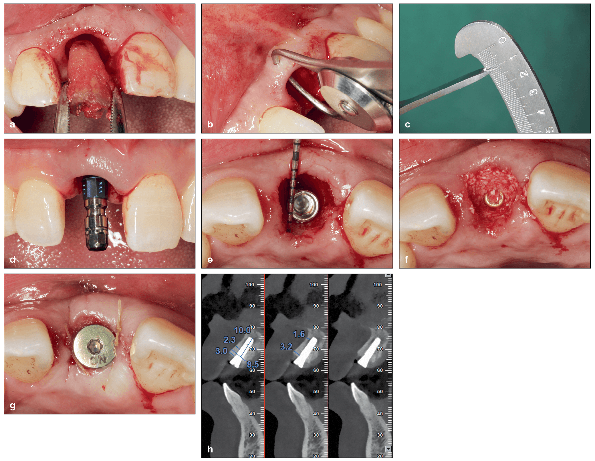
Immediate implant placement in the esthetic zone using a xenograft (DBBM) to pack the gap. (a) Minimally traumatic tooth extraction of a fractured maxillary right central incisor. (b and c) The buccopalatal width of the buccal bone was measured at approximately 1 mm. (d) Implant placement. (e) The implant was placed palatally in the correct 3D position. A buccal gap was created following implant placement. (f) The buccal gap is filled with DBBM. (g) A cover screw is placed with a collagen sponge. (h) Final CBCT after implant placement demonstrating adequate buccal bone. (Case performed by Dr Yufeng Zhang.)

Intended for a global audience and with a watchful eye on the future, the book’s contents are not limited to what is currently and widely commercially available. That’s where the “next-generation” aspect comes in: in addition to gold-standard materials and other available biomaterials, the book also dedicates ample space to new and emerging biomaterials that, while not available everywhere today, are certainly exciting and something clinicians should be aware of for the near future.

Dr Miron provides a few examples. “Today in Europe, they’re using a lot of hyaluronic acid for periodontal regeneration. There are a lot of benefits to that product, and it’s being heavily utilized and studied in Europe, but it’s not being used at all today in the United States. Nobody’s talking about it, nobody’s really doing any experiments with it, and it’s not used even in university settings. In Japan they’re doing great things with atelocollagen right now, which can be processed from the collagen components within the bone structure to create a less-immunogenic bone graft material that favors less of a foreign body reaction. We need to be learning about that here in North America, and most clinicians have not even heard of it! We wanted to collaborate with all of these different colleagues around the world and in such an internationally minded book to allow for more widespread sharing of knowledge.”

Up and Coming: Biomaterials to Watch

With a large mix of biomaterials covered ranging from those still in various stages of development and those clinicians can put to use today, it begs the question: Which of these next-generation biomaterials will become the new standard in coming years?

“The whole concept of this book,” Dr Miron explains, “is that we wanted to develop a textbook that was simultaneously looking at the present and to the future. In this edition of the book, the biomaterials that are just being launched right now and are treated in the text as ‘next-generation biomaterials’ have a lot of preclinical data but not a lot of clinical data. Our plan is to update the book in 5 to 8 years with new biomaterials. By then, there will be a lot more clinical data for many of these biomaterials that are considered new in 2019, and the biomaterials that are considered future today may very well be the standard of care in 5 to 10 years. And by that time, there will certainly be new and exciting developments of next-generation biomaterials, and the cycle will continue with each edition of the book. It should be an exciting project to work on for years to come.”

So taking into account the long R&D gestation period and the long-shot statistics of commercial success for each biomaterial, which next-generation biomaterials from this book would Dr Miron place his bet on becoming the new standard in the years to come?

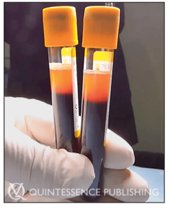
The newer formulation of liquid PRF found in the top 1-mL layer of centrifugation tubes following a 700-rpm spin for 3 minutes. This liquid can be collected in a syringe and reinjected into defect sites or mixed with biomaterials to improve their bioactive properties.

“I think what will grow the fastest and are the most exciting are the more natural biomaterials,” Dr Miron states. “I think that the dentin grinder from chapter 8 of the book—where you actually take the tooth that you just extracted from the patient and grind it up and use it as a dentin particulate graft —is going to grow tremendously over the next 5 to 10 years. I also think that things like platelet concentrates are still under-studied today, and further improvements are necessary going forward. We need to better understand the regenerative potential of each of these biomaterials. One barrier for these natural materials, though, is that they are derived cheaply from the human body. Unfortunately, we’ve learned that the cheaper a biomaterial is, the less commercial involvement they attract. For example, if you extract a tooth and grind it right in the clinic to make a bone graft, your cost is very low. It’s the same with platelet concentrates where you use the patient’s own blood and concentrate growth factors following centrifugation. The problem and downfall with these more ‘natural’ materials is that there isn’t enough money made for companies to support further research and improvement of these biomaterials owing to a general lack of funding. Unfortunately, these materials are more biologic, easier to obtain, and, I would say, probably healthier for you, and they aren’t being studied enough to be viable commercially. And that is a real disadvantage for the field and for patients because they could benefit tremendously from these more natural materials. This is where I think the National Institutes of Health (NIH) should get involved with funding some of these projects, since I do believe it is in the best interest of the national health of our patients.”

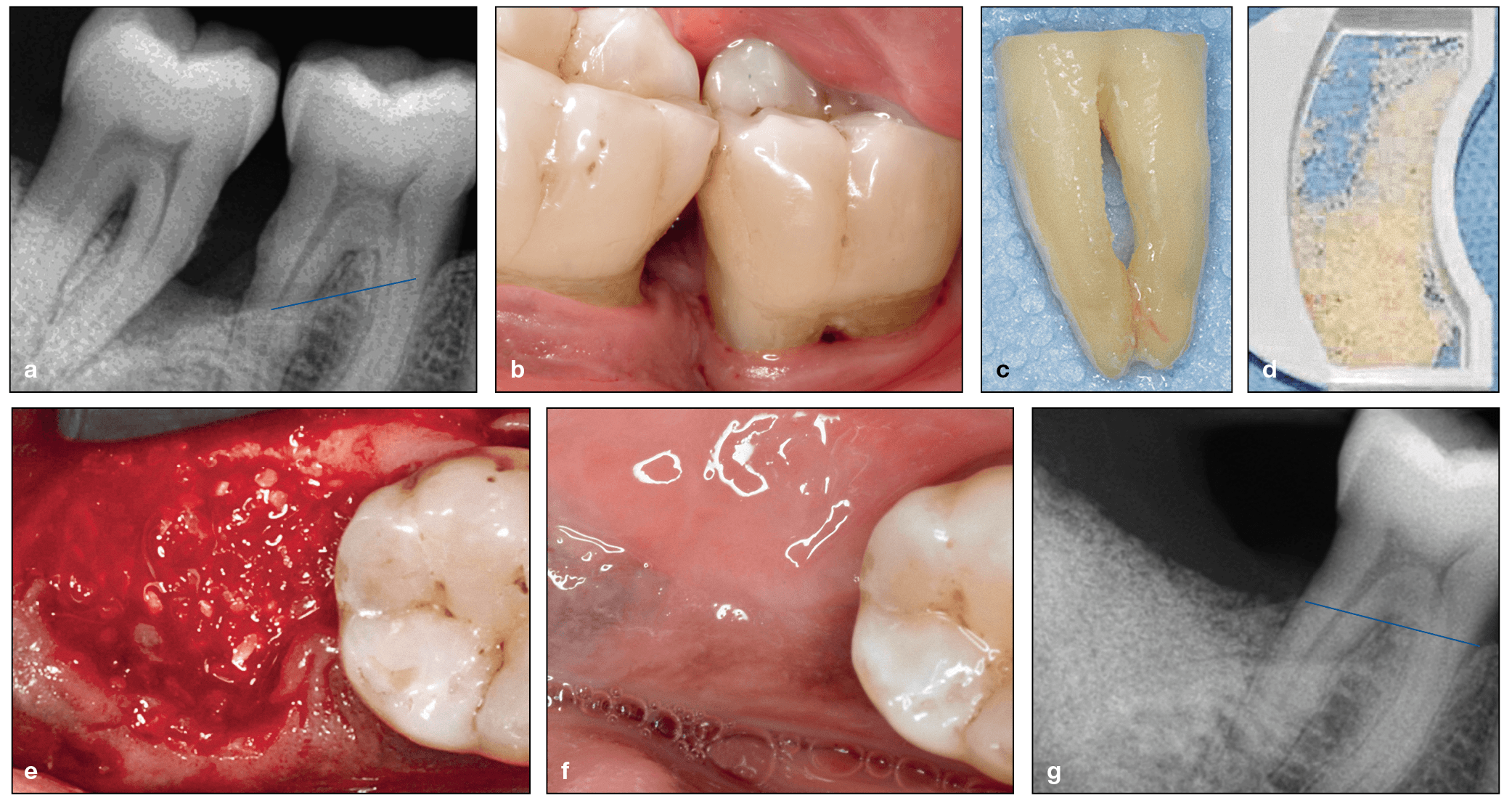
(a) Radiograph demonstrating progressive bone loss of the mandibular left first and second molars, which are periodontally involved. (b) Clinical view of the intrabony defects and exposure of the furcation. (c) The second molar was extracted, and the roots were cleaned. (d) Particulate was prepared using the Smart Dentin Grinder. (e) The particulate dentin was utilized in the extraction socket and grafted against the distal exposed roots of the first molar. Observe the blood that adsorbed onto the dentin particulate graft. (f) Six weeks postextraction, a clinical site next to the first molar shows healthy mucosa. (g) Three months after surgery, the dentin graft is stable within the extraction socket and there is better periodontal support around the first molar. (Case performed by Dr Justin Cifuni.)

Despite the issues these low-profit biomaterials face in gaining wider commercialization, they certainly connect with clinicians.

“The more clinicians become aware of these products, the more they use them, and the more they see a benefit and more interest they create.” Dr Miron explains. “I was in a course taught by Michael Pikos, and he referred to this as ‘going green.’ I laughed so much when I heard him say that because it’s a good way to phrase it: it’s a more organic, more natural way to do things. The advantage to the patient is that you don’t necessarily need to introduce a foreign body material into their body. You don’t have to take a collagen source from the pericardium of a cadaver or the tendon of an animal. You can actually accomplish a lot naturally. I think that this avenue of future research is really exciting, and I think if we can make biomaterials healthier in general for patients, that would be great for our profession. There are certainly indications where you absolutely require a collagen membrane, without question, just like there are indications where you absolutely need an allograft. But whenever we can do things more naturally, it’s less expensive, and if it leads to similar or better outcomes for our patients, then why not?”

While more natural materials provide a marked cost advantage, Dr Miron insists there are other advantages to biomaterials that may cost more, which emphasizes that the greatest benefit lies in having options. “In the book,” he explains, “we also discuss options for biomaterials related to cost. As the field progresses, materials are typically going down in cost. There are still some, especially with regard to growth factors, that are quite expensive, and a perfect example of that is rhBMP-2, which is sold by Medtronic and costs upwards of $600 per case. Now the argument for rhBMP-2 that a lot of expert clinicians such as Robert Marx are making, is that in some indications you can completely replace the need to harvest autogenous bone from the patient’s body by instead using rhBMP-2. You could maybe replace having to get bone from the iliac crest or the tibia and instead use growth factors. You can then present both options to your patient. You’ll notice that in a lot of leading hospitals, and in oral and maxillofacial surgery especially, they’re trying to minimize morbidity of the patient by utilizing some of these newer technologies when it comes to growth factors and recombinant proteins. A lot of patients will go down that route because it makes a lot of sense: pay a little more but avoid a second surgical site with additional morbidity. The good thing is that the more we utilize some of these biomaterials, the lower the costs typically become. I truly believe that the future of medicine in general—not just dentistry—really relates to growth factor use. This is one area that every clinician should be aware of and keeping tabs on.”

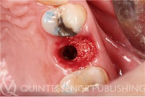
Clinical photograph demonstrating the use of Osopia to fill the buccal gap during immediate implant placement. Because Osopia is both mineralized and osteoinductive, it offers the advantage of inducing rapid new bone formation while resorbing more slowly over time when compared to DFDBA. (Case performed by Dr Albert Barroso Panella.)

But there are plenty more biomaterials in the book worth getting excited about. “Another avenue that I personally find very exciting are new synthetic bone graft materials that are now osteoinductive, meaning the bone graft material is able to form ectopic bone. A simpler explanation is that these materials are kind of super inducers of bone regeneration. Today there are only two FDA-approved osteoinductive biomaterials: a demineralized allograft coming from another human cadaver or rhBMP-2, which is very expensive. Recently, however, a group of researchers in the Netherlands found a way to make osteoinductive synthetic materials. It’s the first time anyone’s ever been able to do that, and we’ve included their research in this book. This material is being used clinically in Europe, and it currently presents itself as being a big breakthrough. It’s already FDA-approved for orthopedic use here in USA, and I’m assuming that it will be approved for dental use very shortly, and it’s going to have a huge impact on the field.”

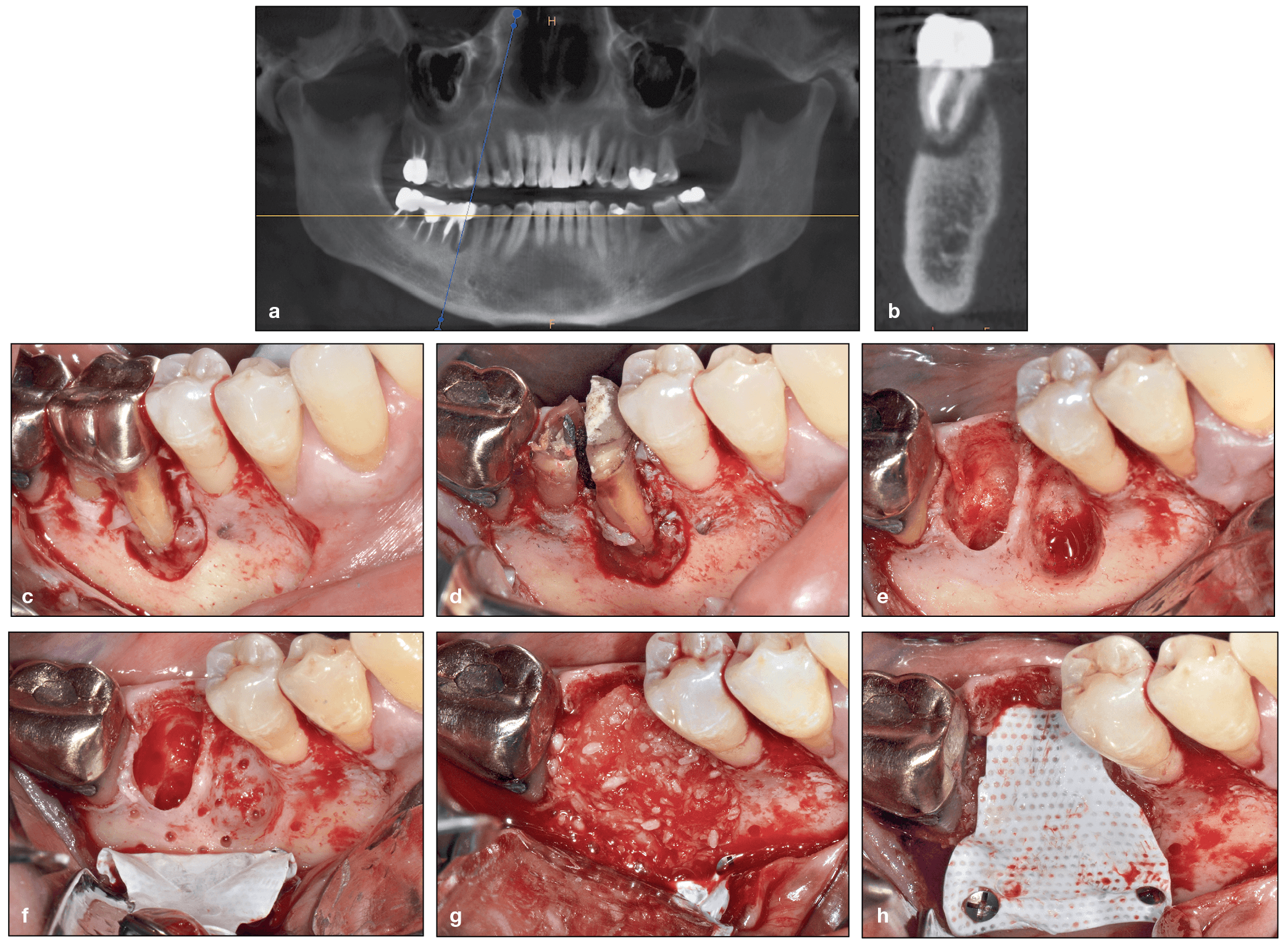
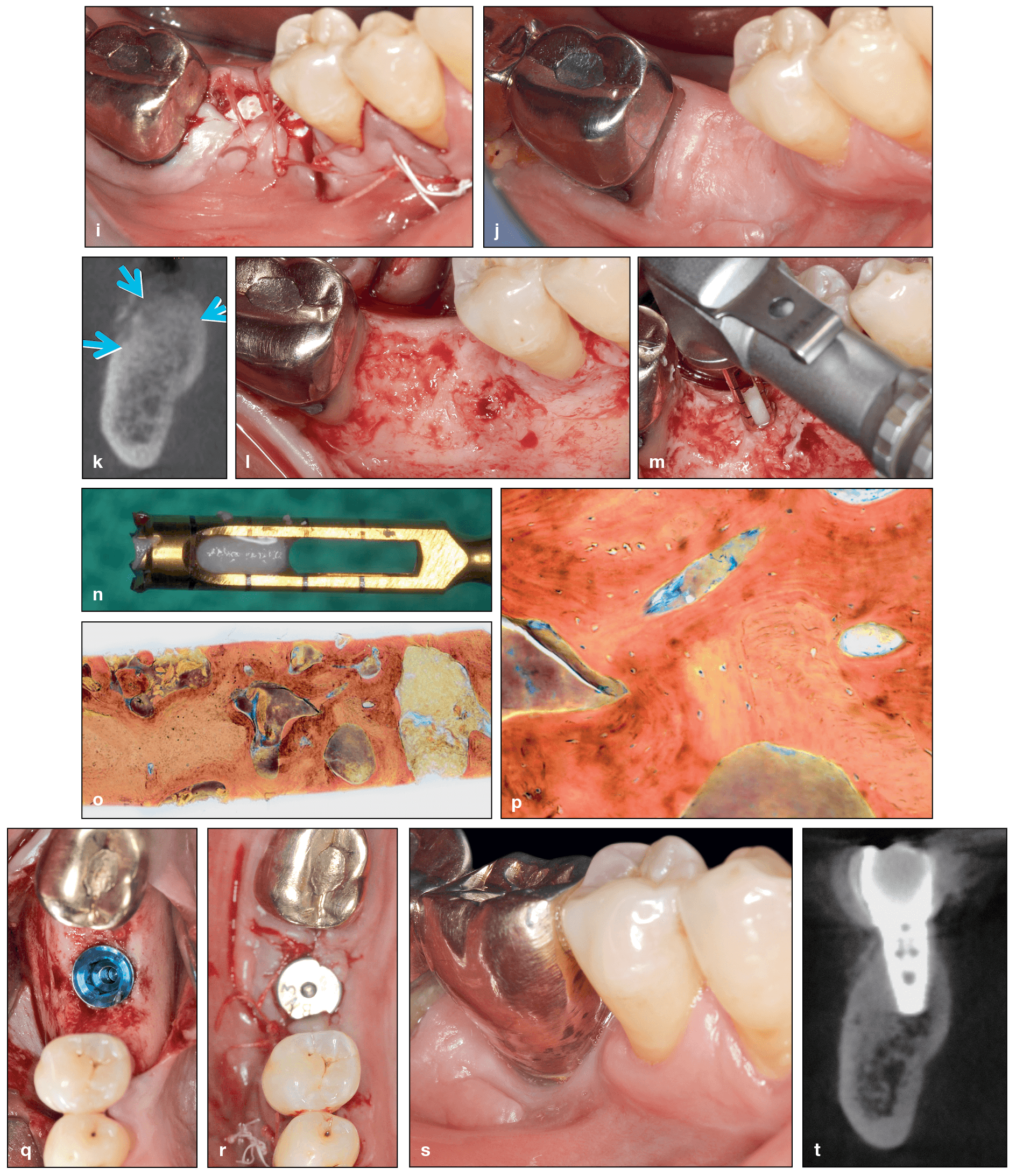
Use of rhBMP-2 following extraction of a mandibular first molar with exposed root surfaces. (a and b) CBCT demonstrating a periapical lesion at the site of the mandibular right first molar and complete loss of the buccal plate. (c) The extent of bone loss is observed after flap elevation. (d and e) Extraction of the mandibular first molar. Note that the tooth was split in half and removed atraumatically. (f) After decortication and intramarrow penetration, a titanium-reinforced dense polytetrafluoroethylene (dPTFE) membrane was fixed apically. (g and h) rhBMP-2 was mixed with an allograft, and the composite graft was used to fill the bone void, after which the membrane was secured.

(cont) (i) Final sutures. (j and k) At 7 months postoperative, the ridge was maintained in both the horizontal and vertical dimensions. (l to n) A flap was raised, and a bone core sample was harvested. (o and p) Histology demonstrating excellent new bone formation with viable osteocytes following a 7-month healing period. (q and r) Implant placement. (s) Final restoration at 5 years. (t) CBCT at 5 years demonstrating excellent bone maintenance, especially on the buccal contour of the implant. (Case performed by Dr Michael A. Pikos.)

“I have also done a lot of research with rhBMP-9,” Dr Miron continues, “and that research is very fascinating. What happens with rhBMP-9 versus rhBMP-2, which is the current standard, is that you can use 5 to 20 times less rhBMP-9, and it’s more effective. The advantage then is that you’re giving your patients less of a medication or drug, which is always better, and it should theoretically cost less because you’re using less growth factor. One other area I find presents itself as having huge potential is all the work that has been done with Tetranite, a novel bone adhesive that is still in the preclinical testing phase. It’s basically a bone glue that is very biocompatible, and it allows you to place an implant and obtain primary stability where previously you may not have been able to place it. Tetranite adheres both to the implant metal as well as to bone and provides primary stability: ISQ values are excellent, they remain high over time, and it’s fully resorbable and replaced with native bone within a year, making it an absolutely dream product for dentistry if all the data holds up during clinical testing.”

(a to f) Clinical photographs of a canine alveolar ridge that was preserved by injecting Tetranite into the extraction sockets. (g to i) Postoperative healing at 2 weeks (g), 6 weeks (h), and 9 weeks (i). Note the uneventful healing of the soft tissues and the overgrowth of the exposed material at week 2 (g). (j) Exposed ridge prior to osteotomy preparation at 12 weeks. (k and l) Clinical views 3 weeks and 9 weeks after implant placement, respectively. (m) CBCT image of the implants 26 weeks after injection of the Tetranite and 14 weeks after implant placement. The posterior implant is successfully osseointegrated, while the anterior site is too shallow for a successful outcome.

(a) Harvested tissue at week 26 demonstrates that TN integrates into host bone around implants even when β-TCP granules are added (ground section, trichrome stain). (b and c) Higher-magnification views of the areas indicated by the rectangles in a. After 26 weeks, TN is fully biocompatible and integrates into host bone. Furthermore, resorption of the material is observed with visual bone-to-implant contact. I, implant; nb, new bone; G, graft; bb, bony bridge; g, gingival tissue; lb, lamellar bone.

The Future Is Now: Moving Forward

The sheer number of all of these new and exciting biomaterials can no doubt feel overwhelming for the clinician who just wants to better treat their patients. But Dr Miron, who also teaches courses on these biomaterials, knows exactly how to guide these clinicians through the unfamiliar.

“In my experience, clinicians in general don’t just want to be told which material to use. They actually enjoy understanding how and why to use each material, why they’re used with different protocols, why you need BMPs here, and why you need a xenograft there. So with this book we really just wanted to answer the why, why, why. Why is Istvan Urban using autogenous bone with xenografts? Why is Daniel Buser doing contour augmentation? Why is Anton Sculean using hyaluronic acid? Why are Michael Pikos and Robert Marx using BMPs? There are real biologic reasons for using different biomaterials in different clinical indications. At the same time, we wanted to write it in a way that would feel more like a general story that you can read through with cases from a number of expert clinicians who have contributed, and the information just flows naturally. The science is there because Yufeng Zhang and I have extensively investigated each of these biomaterials, but the clinical practicality is also there throughout because we are also clinicians and, more importantly, because we collaborated on this project with colleagues whom I consider to be the top clinicians from around the world.”

No one can save every single tooth. But with better biomaterials and appropriate selection, we can surely get closer to achieving these idea results.

Dr Richard J. Miron
Dr Miron also has a message for the clinicians who are comfortable with their selection of biomaterials and the results they obtain from them. “There are always ways to achieve better results. No one can save every single tooth. No one can predictably augment 8 mm of bone vertically. But with better biomaterials and appropriate selection, we can surely get closer to achieving these ideal results. The goal is not to say that what you’re doing is wrong, could be improved, or that you should do it another way, it’s just about educating colleagues so they are aware of what’s out there. This book is a highlight of what’s available today and what’s coming tomorrow. The reality is that the majority of practicing clinicians have never heard of 50% of the biomaterials we are presenting in this book, and it will be eye-opening for a lot of dentists to see how the field is actually advancing and what great things will be available in the near future.”

Similar to the sheer number of biomaterials introduced is the staggering amount of “information” about them, and that can be a problem for clinicians who are trying to understand the full picture about their biomaterial options.

Clinicians don’t want nonsense anymore.

Dr Richard J. Miron
“One of the most shocking things I learned from my experience working and studying in Europe is the big push on marketing here in North America as compared with Europe. There is a lot less marketing for biomaterials in Europe, whereas here, when you walk around a big conference, there are a ton of sales reps who are coming to you and telling you what you should use. Most clinicians are actually getting their research information from a sales rep, and that is really frustrating to me. There should be more sources of better information from an academic point of view that clinicians can access, which was a huge part of why we wrote this book. Clinicians don’t want nonsense anymore. Dentists should have access to everything that’s out there so they can figure out what they want to utilize for their own practices based on their own personal preferences and based on sound scientific evidence. I think from this point of view, clinicians will respond well to the book, and I hope that this book will change the way we learn and teach about these biomaterials.”

Next-Generation Biomaterials for Bone & Periodontal Regeneration will no doubt open a new door into the future for clinicians worldwide. But just like no implant can guarantee success without the hands of a skilled surgeon, no biomaterial alone can guarantee successful bone or periodontal regeneration. A successful outcome will always rely on a skilled and knowledgeable clinician. What this book will do is increase the depth of the reader’s knowledge base and enable them to make more informed and evidence-based decisions about their biomaterials selection depending on the case, treatment, surgery, and their patient.


Dr Richard J. Miron is currently Visiting Faculty in the Department of Periodontology at the University of Bern in Switzerland, where he completed his PhD studies. He has published over 150 peer-reviewed articles and lectures internationally on many topics relating to growth factors, bone biomaterials, and guided bone regeneration. Dr Miron has been awarded many top international prizes in regenerative dentistry and implant dentistry, including the ITI André Schroeder Research Prize, the International Association for Dental Research Young Investigator of the Year award in the field of implant dentistry, and the American Academy of Implant Dentistry Young Investigator grant award. He has completed postdoctoral research fellowships in Switzerland, Canada, China, Spain, and the United States.


Dr Yufeng Zhang is the Luojiashan Distinguished Professor and chief physician at the Wuhan University Medical and Dental School in Wuhan, China. He completed a two-year peri-implant bone biology research fellowship at Queensland University of Technology in Brisbane, Australia, and in 2010 he completed an ITI scholar position at the University of Bern in Switzerland. His research is primarily engaged in the field of implanted biomaterials used for alveolar bone and periodontal regeneration. Dr Zhang was one of the first researchers to propose the concept of gene-activated tissue engineering scaffolds, developing a scaffold-gene integration system that has its own biologic activity in vivo due to the autocrine growth factor. He has since been responsible for the development of several biomaterials with numerous industrial partners.


Next-Generation Biomaterials for Bone & Periodontal Regeneration

Edited by Richard J. Miron and Yufeng Zhang

New and innovative biomaterials are being discovered or created in laboratories at an unprecedented rate, but many of them remain entirely foreign to practicing clinicians. This book addresses this gap in knowledge by summarizing some of the groundbreaking research performed to date on this topic and providing case examples of these biomaterials at work. The book begins with a review of the biologic background and applications of bone grafting materials utilized in dentistry. The principles of guided tissue and bone regeneration are covered in detail, including many recent advancements in barrier membrane technologies as well as use of platelet-rich fibrin and various growth factors, and many next-generation materials that will optimize future bone and periodontal regeneration are presented. The final chapter is designed to help clinicians select appropriate biomaterials for each specific regenerative protocol. Much like one implant size and shape cannot be utilized for every indication in implant dentistry, one bone grafting material, barrier membrane, or growth factor cannot maximize regenerative outcomes in all clinical situations. This textbook teaches clinicians how to utilize biomaterials in an appropriate, predictable, and evidence-based manner.

384 pp; 960 illus; ©2019; ISBN 978-0-86715-796-3 (B7963); $218


This article was written by Caitlin Davis, Quintessence Publishing.

©2019 BY QUINTESSENCE PUBLISHING CO, INC. PRINTING OF THIS DOCUMENT IS RESTRICTED TO PERSONAL USE ONLY. NO PART MAY BE REPRODUCED OR TRANSMITTED IN ANY FORM WITHOUT WRITTEN PERMISSION FROM THE PUBLISHER.

Posted in Books, Feature, Implant Dentistry, Multidisciplinary, Periodontics | Tagged , , , | 4 Comments

Introducing The Sinus Bone Graft, Third Edition, edited by Ole T. Jensen
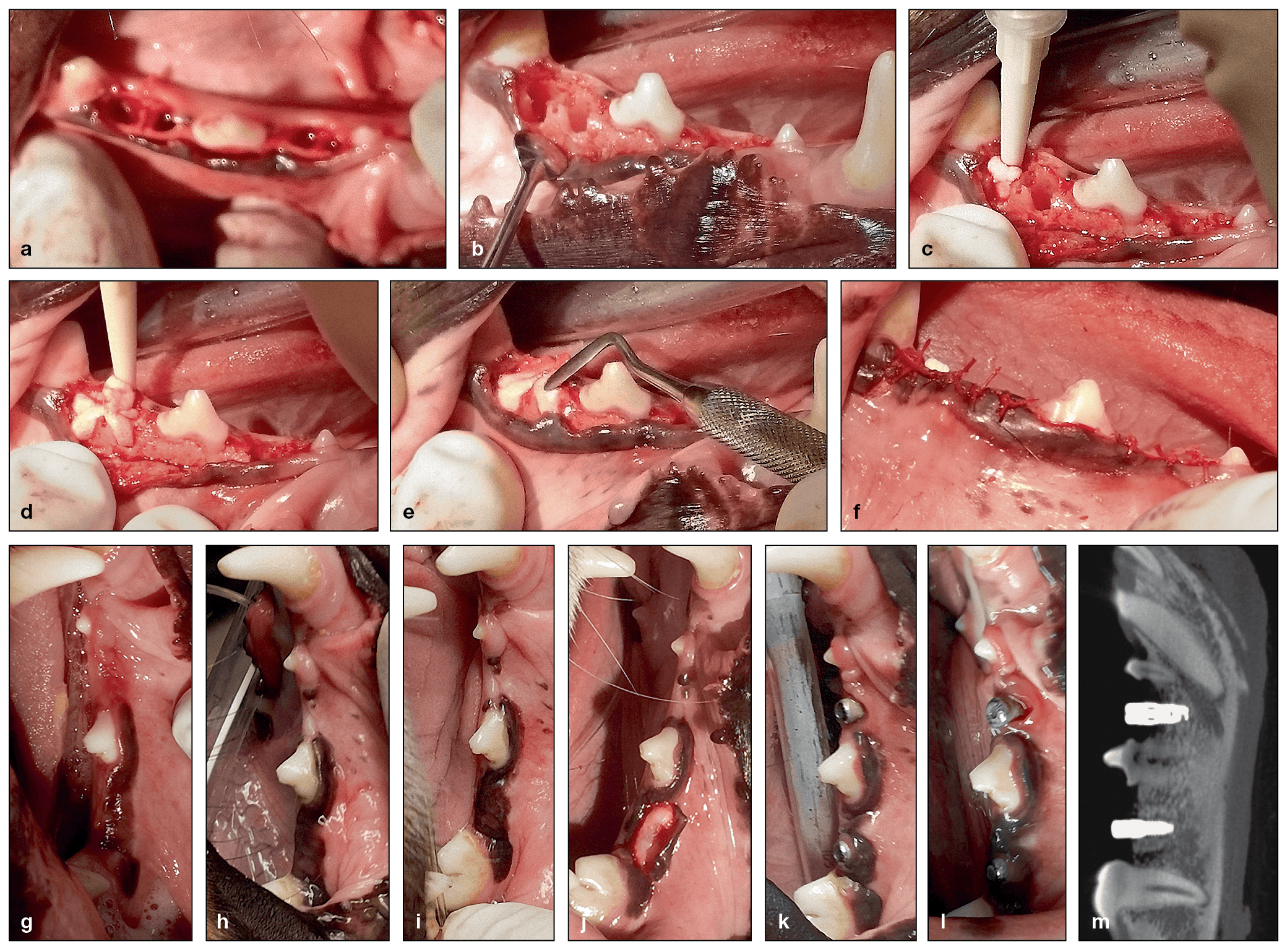
Reading time: 2 minutes

As research proceeds on treatment of the resorbed posterior maxilla, new techniques and innovations continue to be adopted to solve this clinical problem. While the previous edition of The Sinus Bone Graft provided detailed information on the types of grafting materials and procedures available at the time, this completely revised version looks to the future with new strategies for treatment, some of which avoid grafting altogether. Learn more in this Introduction to the book by Hilt Tatum Jr, DDS.
 

Jensen Intro


The Sinus Bone Graft, Third Edition

Edited by Ole T. Jensen

“This excellent updated volume describes the latest advances and modifications of the sinus elevation procedure, including alternatives such as corrective osteotomies, zygomatic implants, and even graftless treatment.”

—Hilt Tatum, Jr


This book not only reviews the time-tested lateral window approach for sinus elevation and grafting but also describes a variety of techniques to approach the sinus transcrestally with or without grafting material. One section of the book is devoted entirely to the different types of implants and implant placement techniques available, and there are many clinical case studies and descriptions of how to perform specific surgical procedures.

288 pp; 948 illus; ©2019; ISBN 978-0-86715-791-8 (B7918); US $168

Posted in Books, Implant Dentistry, Oral and Maxillofacial Surgery, What's New | 3 Comments