Author Spotlight: Daniel M. Laskin

Reading time: 12 minutes

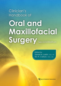
Few names in modern oral and maxillofacial surgery are as respected as that of Daniel M. Laskin, DDS, MS. Throughout his career, Dr Laskin has made significant contributions to the specialty through his research, literature, and teaching and has also contributed immensely to the advancement of the specialty through his involvement in professional organizations. To commemorate the recent publication of the second edition of the Clinician’s Handbook of Oral and Maxillofacial Surgery, we offer this Author Spotlight in celebration of Dr Laskin’s incredible career and in gratitude for his contributions to our own body of publications.

A Legacy of Excellence

Temporomandibular Disorders: An Evidence-Based Approach to Diagnosis and Treatment, edited by Daniel M. Laskin, Charles S. Greene, and William L. Hylander (Quintessence, 2006)

Dr Laskin received his dental degree from the Indiana University School of Dentistry in 1947. He then completed a 1-year internship in oral surgery at the Jersey City Medical Center in Jersey City, New Jersey, in 1948 and his postgraduate studies in oral surgery at the University of Illinois at Chicago (UIC) College of Dentistry in Chicago, Illinois, in 1950. In 1951 he completed a master of science in oral pathology at UIC and a 1-year residency in oral surgery at the John H. Stroger Jr. Hospital of Cook County in Chicago, Illinois.

Dr Laskin remained at the UIC College of Dentistry for more than three decades, departing in 1983 as Professor Emeritus. During his tenure there, Dr Laskin taught both in the Department of Oral and Maxillofacial Surgery of the College of Dentistry, where he served as Head of Department from 1973 to 1983, and in the Department of Surgery of the College of Medicine, where he served as Director of the Postgraduate and Graduate Program for the Department of Oral and Maxillofacial Surgery from 1955 to 1983. He also served as Director of the UIC Temporomandibular Joint and Facial Pain Research Center from 1963 to 1983 and as Director of the Oral Surgery Residency Training Program at John H. Stroger Jr. Hospital of Cook County.
Decision Making in Oral and Maxillofacial Surgery, edited by Daniel M. Laskin and Omar A. Abubaker (Quintessence, 2007)

In 1984, Dr Laskin made the move from Chicago to Richmond, Virginia, to serve as Professor and Chairman of the Department of Oral and Maxillofacial Surgery at the Virginia Commonwealth University (VCU) School of Dentistry and of the Division of Oral and Maxillofacial Surgery of the Department of Surgery at the VCU School of Medicine. He directed the VCU Temporomandibular Joint and Facial Pain Research Center from 1984 to 2002. He achieved Chairman Emeritus status from the VCU School of Dentistry in 2002, where he remains involved and continues to lecture.
Treatment of TMDs: Bridging the Gap Between Advances in Research and Clinical Patient Management, edited by Charles S. Greene and Daniel M. Laskin (Quintessence, 2013)

“Dr Daniel Laskin is the quintessential oral and maxillofacial surgery educator,” says Eric Carlson, DMD, MD, EdM, coeditor of the Clinician’s Handbook of Oral and Maxillofacial Surgery, Second Edition. “He has devoted his entire career to education and has impacted every oral and maxillofacial surgeon over the past 50 years of our specialty’s existence. In short, Dan Laskin is synonymous with oral and maxillofacial surgery, and our entire specialty owes him a debt of gratitude. In paying homage to Dr Laskin, I simultaneously acknowledge all oral and maxillofacial surgery educators who are like him in some small way.”

Over the course of his career, Dr Laskin has also served the specialty through his involvement in various national and international professional organizations. He has served as President of the American Association of Oral and Maxillofacial Surgeons (1976–1977), the International Association of Oral and Maxillofacial Surgeons (1983–1986), and the National Academies of Practice (2002–2004). He is a fellow in Dental Surgery of the Royal College of Surgeons of England and the Royal College of Physicians and Surgeons of Glasgow (Hon). He is also a fellow of the American College of Dentists, the American Association for the Advancement of Sciences, the International College of Dentists, and the American Dental Society of Anesthesiology. The list of honors and awards he has received runs two pages long and includes the American Association of Oral and Maxillofacial Surgeons Research Recognition Award in 1978, the William J. Gies Oral Surgery Award in 1979, the American Dental Society of Anesthesiology Heidbrink Award in 1983, and the Donald B. Osbon Award for Outstanding Educator in 1991.
Oral and Maxillofacial Surgery Review: A Study Guide, edited by Din Lam and Daniel M. Laskin (Quintessence, 2015)

Dr Laskin’s research throughout his career has focused on the fields of connective tissue physiology and pathology, craniofacial growth, and the temporomandibular joint and its disorders. He has authored or coauthored more than 900 publications in OMFS and dental research and has won the William J. Gies Editorial Award 8 times with 10 Honorable Mentions. He is the author or coauthor of 18 books, including 6 with Quintessence Publishing.

The Book: Clinician’s Handbook of Oral and Maxillofacial Surgery, Second Edition
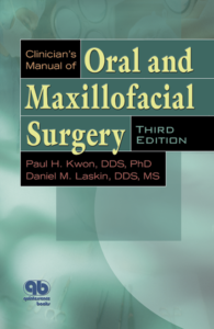
The Clinician’s Manual of Oral and Maxillofacial Surgery, edited by Paul H. Kwon and Daniel M. Laskin (Quintessence, 2001)

The Clinician’s Handbook of Oral and Maxillofacial Surgery was originally adapted from the Clinician’s Manual of Oral and Maxillofacial Surgery, which was edited by Dr Paul H. Kwon and Dr Laskin and saw three editions published between 1991 and 2001.

“The first edition of the Clinician’s Handbook of Oral and Maxillofacial Surgery,” Dr Laskin explains, “was designed to be portable in order to make the information immediately available. However, the amount of information necessary to include made it impossible to accomplish that goal. Therefore, the decision was made for the second edition to further increase the size so that more chapters, illustrations, and summary tables could be included, adding to the usefulness of the book. Condensing easy-to-access, pertinent information for the entire scope of the specialty is an impossible task for one person. However, selecting contributing authors with particular expertise in the various areas helped us achieve that goal and provide the information in a concise, organized manner.”

“As editors of this book, we endorsed individuality for all authors to format their chapters to be practically useful to our readership,” Dr Carlson adds. “We did not insist on a uniform presentation of the chapters, but rather gave authors permission to autonomously format their chapters to make high-yield information readily apparent to the readership in the best interests of proper patient care.”

The updates to both the format and the scope of this second edition contribute to the usefulness of the book as an important clinical resource by putting more of what the practicing oral and maxillofacial surgeon needs into one convenient reference source. It solves the issue of the time-consuming search for a current, reputable, scientifically proven solution for an immediate clinical problem. New chapters have been added on implantology, cleft lip and palate, maxillofacial reconstruction, oral squamous cell carcinoma, and cosmetic surgery.
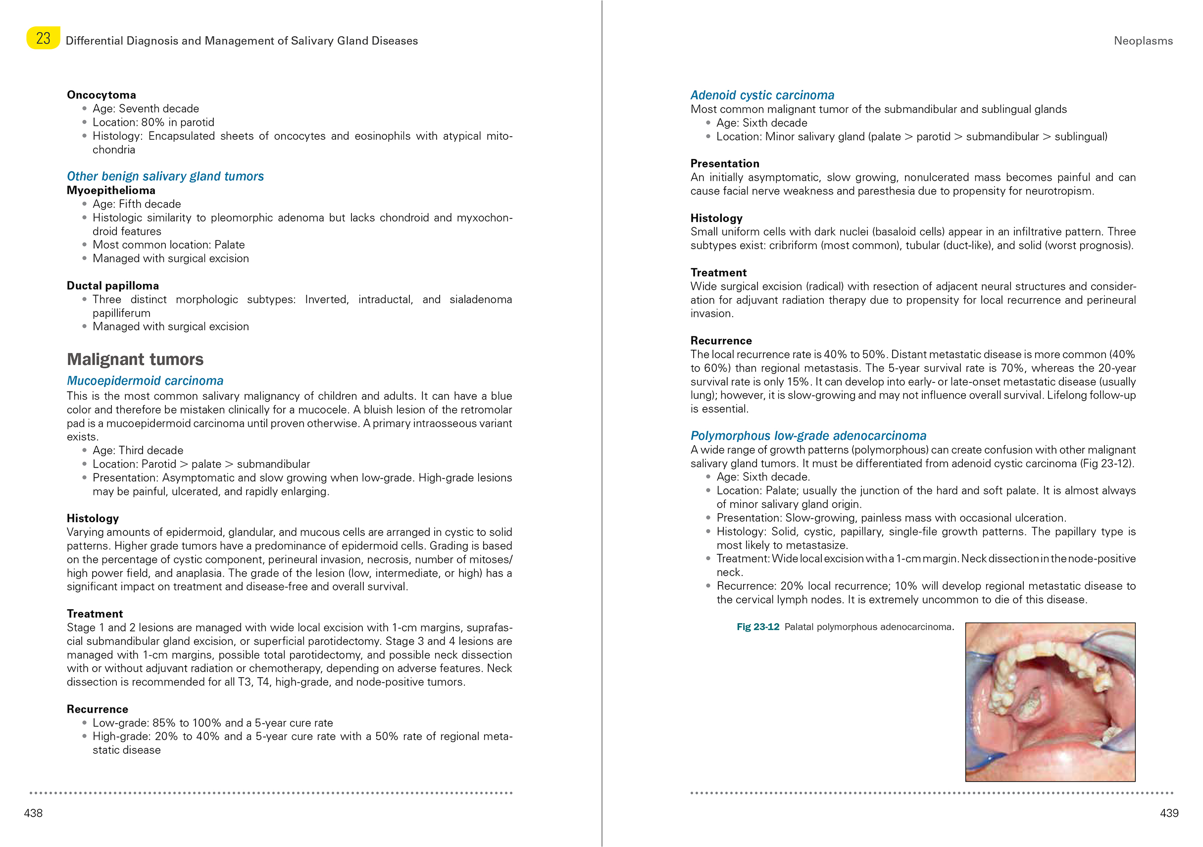
An example page spread from the expanded second edition of the Clinician’s Handbook of Oral and Maxillofacial Surgery.

“I have produced two types of books,” Dr Laskin says. “I have edited comprehensive textbooks on temporomandibular disorders that bring together experts to provide detailed information on one specific subject, and then there are those like this book that gathers and summarizes information from the entire specialty—pieces that would ordinarily be found in various different sources—into one site for quick reference. Although both types of books serve different purposes, in totality they provide the necessary information for practicing clinicians as well as residents to remain current in the specialty.”

The Clinician’s Handbook of Oral and Maxillofacial Surgery, Second Edition is not intended to replace the exhaustive textbook; instead, it synthesizes the information learned in postgraduate study and residency into a practical, clinically accessible format that clinicians can read cover to cover to review and familiarize themselves with the organization of information, then consult quickly for on-the-spot questions and answers as well as protocol validation.
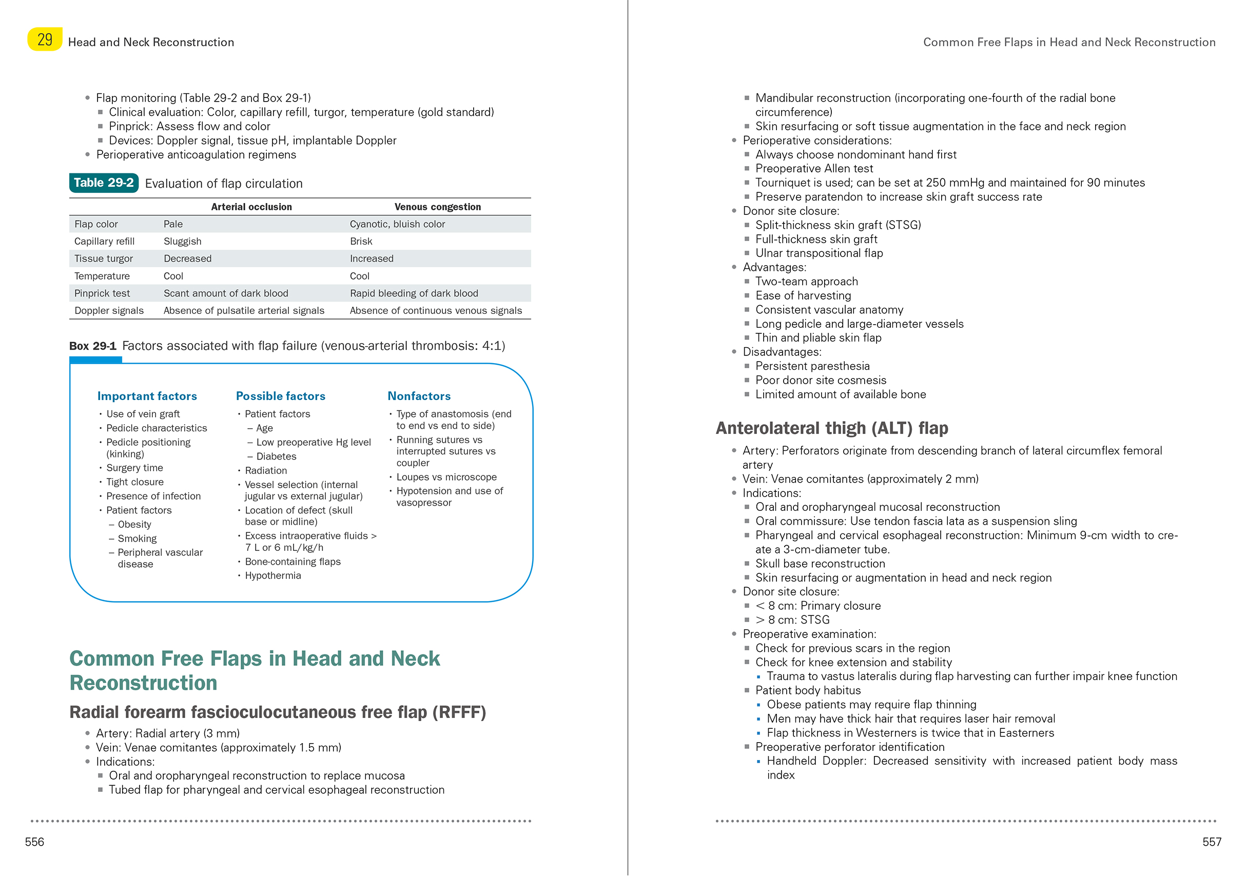
An example page spread from the expanded second edition of the Clinician’s Handbook of Oral and Maxillofacial Surgery.

“The popularity of this type of book,” Dr Laskin observes, “is indicated by the fact that this book ran for three editions in its original format and is now entering its second edition in the new format. For clinicians, this book still serves as a quick way to gain information in one place that ordinarily might be contained in various sources. For residents, it serves a similar purpose, as well as a good resource in preparation for the American Board of Oral and Maxillofacial Surgery.”

With the age of this book in both of its formats currently standing at just 27 years—young compared with many long-running textbooks—it is important to consider the future for a book with this level of impact and reputation, something Dr Laskin has certainly kept in mind while planning this second edition. In addition to expanding the format to increase the scope and impact of the book, he also brought in Dr Carlson as his coeditor.

“Because I am approaching the end of my career,” Dr Laskin says, “I made the decision to include a coeditor with outstanding academic and clinical credentials on this edition so that this publication will continue to be available in the future.”

“It is particularly meaningful to me,” Dr Carlson says, “that—at the age of 94—Dr Laskin continues to make outstanding contributions to our specialty’s literature. I feel honored to have served as Dr Laskin’s coeditor of this very important work. In many respects, peer-reviewed publications have supplanted textbooks in terms of their ability to provide current and meaningful evidence-based information for educational and patient-care purposes. However, textbooks and handbooks like this title go further than peer-reviewed publications can go by gathering a large body of current research, clinical results, and expert analysis into a comprehensive and cohesive reference source. Based on these realizations, it is always essential to plan the issue of a second edition of an important textbook. The information contained within the second edition is in keeping with providing current and meaningful information to trainees and practicing surgeons in our specialty. Furthermore, the incorporation of high-quality color images and illustrations enhances the educational value of the written information. I anticipate that the readership will peruse this textbook in preparation for challenging patient-care scenarios to ensure their adherence to current best practices.”

Reflections of Gratitude

As Dr Laskin reflects on his career, we reflect on the impact of his contributions to the field as well as our relationship with him as his publisher. It has been our pleasure to publish six books with Dr Laskin, and we have come to appreciate him as an editor with a well-developed vision for all of his publications, a reliable sense of attention to detail, and the clear communication skills necessary to coordinate a large group of contributors. In each project we have done with him, he has provided strong guidance and an incomparable source of knowledge and feedback while we have done our best to bring his visions to life on the page.

“Unlike with the conglomerate publishers,” Dr Laskin says, “working with Quintessence is like working with a family. You receive personal attention, you get to know the people with whom you work, and everyone’s goal is to produce an outstanding publication.”

Some people have a curriculum vitae so impressive that outstanding becomes the result the rest of us come to expect from that person because it has been achieved so many times before, and Dr Laskin is a true exemplar of this. It has been our pleasure to be his publisher, and we offer him our sincerest gratitude for the indelible effect he has had on the field of oral and maxillofacial surgery as well as on our world of dental publishing.


Daniel M. Laskin, DDS, MS, has been a major contributor to the field of oral and maxillofacial surgery for more than 50 years. He received his dental degree from the Indiana University School of Dentistry in Indianapolis, Indiana, and his master of science in oral pathology from the University of Illinois at Chicago (UIC) in Chicago, Illinois. He completed his postgraduate studies in oral surgery at UIC and a 1-year residency at the John H. Stroger Jr. Hospital of Cook County in Chicago, Illinois. He served at the UIC College of Dentistry for more than 30 years, departing in 1983 as Professor Emeritus. In 1984, he left Chicago to serve as Professor and Chairman of the Department of Oral and Maxillofacial Surgery at the VCU School of Dentistry and of the Division of Oral and Maxillofacial Surgery of the Department of Surgery at the VCU School of Medicine. He directed the VCU Temporomandibular Joint and Facial Pain Research Center from 1984 to 2002. He achieved Chairman Emeritus status from the VCU School of Dentistry in 2002, where he remains involved and continues to lecture. He has served as President of the American Association of Oral and Maxillofacial Surgeons (1976–1977), the International Association of Oral and Maxillofacial Surgeons (1983–1986), and the National Academies of Practice (2002–2004). He is a fellow in Dental Surgery of the Royal College of Surgeons of England and the Royal College of Physicians and Surgeons of Glasgow (Hon). He is also a fellow of the American College of Dentists, the American Association for the Advancement of Sciences, the International College of Dentists, and the American Dental Society of Anesthesiology. He has served on the editorial board of more than 20 publications, contributed to more than 900 publications in OMFS and dental research, and has authored or co-authored 18 books. Throughout his career, Dr Laskin’s research has focused on connective tissue physiology and pathology, craniofacial growth, and the temporomandibular joint. His contributions to the field and his dedication to dental education have earned him a large number of honors and fellowships, and the impacts of his career will continue to benefit the field of oral and maxillofacial surgery for decades to come.

 

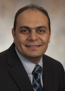
Eric R. Carlson, DMD, MD, EdM, is Professor and Kelly L. Krahwinkel Chairman of the Department of Oral and Maxillofacial Surgery at the University of Tennessee, where he also serves as Director of the Oral and Maxillofacial Surgery Residency Program and the Oral/Head and Neck Oncologic Surgery Fellowship Program. He received his dental degree from the University of Pennsylvania School of Dental Medicine, his medical degree from the University of Miami Leonard M. Miller School of Medicine, and his master of education degree from the Harvard University Graduate School of Education. Dr Carlson is a fellow of the American College of Surgeons and has previously served as Chair of the American Association of Oral and Maxillofacial Surgeons Special Committee on Oral and Maxillofacial Surgery Parameters of Care, as the Oral and Maxillofacial Surgery Commissioner for the Commission on Dental Accreditation (CODA), and as Chair of the CODA Oral and Maxillofacial Surgery Residency Review Committee. He has also served as the Surgical Oncology and Reconstruction Section Editor for the Journal of Oral and Maxillofacial Surgery. Dr Carlson’s clinical expertise is in oral/head and neck tumor surgery, and his research focuses on the molecular aspects of cancer diagnosis and treatment.


Clinician’s Handbook of Oral and Maxillofacial Surgery, Second Edition

Edited by Daniel M. Laskin and Eric R. Carlson

There are frequent situations in which oral and maxillofacial surgeons find themselves in need of an immediate answer to a clinical problem. However, this can involve a time-consuming search for the appropriate reference source. This book continues the format of the previous edition by providing a single place to quickly find information on a diverse range of clinical topics, including dentoalveolar surgery, maxillofacial trauma, craniofacial anomalies, and oral pathology. All of the previous chapters have been updated, and new chapters on implantology, cleft lip and palate, maxillofacial reconstruction, oral squamous cell carcinoma, and cosmetic surgery have been added. Moreover, increasing the size of the book has allowed for the inclusion of many summary charts, tables, clinical photographs, and radiographs, which was not possible in the previous version. As a result, this new edition provides expanded information in an improved format.

Although this book is designed as a quick reference source, familiarizing oneself with its content in advance will both add to the reader’s general knowledge base and improve the ability to find information quickly in urgent situations. Residents in oral and maxillofacial surgery should find its content particularly useful during their clinical training, and the concise organization of the material should also be helpful to them in retaining information when subsequently preparing for the American Board of Oral and Maxillofacial Surgery.

624 pp (softcover); 374 illus; ©2019; ISBN 978-0-86715-730-7 (B7307); $168

Posted in Author Spotlight, Books, Feature, Oral and Maxillofacial Surgery | Tagged , , | Leave a comment

Quintessence Roundup: October

Reading time: 11 minutes

October Monthly Special


Dentistry with a Vision: Building a Rewarding Practice and a Balanced Life

Gerald I. Kendall and Gary S. Wadhwa

Dental practitioners and their staff will find this engaging story an easy and fun way to learn how to implement powerful, scientifically based project-management principles into their practice. It will help you to identify the few key issues that are holding you back; substantially increase your profits within weeks; treat more patients in less time without sacrificing quality of care; reduce waste, repeated work, and stress; improve relationships among the practitioners, staff, and outside service providers; work fewer hours while performing more of the kind of treatments you enjoy; increase referrals and patient demand; and develop an effective scheduling system.

200 pp (softcover); 10 illus; ©2009; ISBN 978-0-86715-489-4 (B4894); $38 Special price! $25


New Titles in Books


Cephalometry in Orthodontics: 2D and 3D

Edited by Katherine Kula and Ahmed Ghoneima

Cephalometrics has been used for decades to diagnose orthodontic problems and evaluate treatment. However, the shift from 2D to 3D radiography has left some orthodontists unsure about how to use this method effectively. This book defines and depicts all cephalometric landmarks on a skull or spine in both 2D and 3D and then identifies them on radiographs. Each major cephalometric analysis is described in detail, and the linear or angular measures are shown pictorially for better understanding. Because many orthodontists pick specific measures from various cephalometric analyses to formulate their own analysis, these measures are organized relative to the skeletal or dental structure and then compared or contrasted relative to diagnosis, growth, and treatment. Cephalometric norms (eg, age, sex, ethnicity) are also discussed relative to treatment and esthetics. The final chapter shows the application of these measures to clinical cases to teach clinicians and students how to use them effectively. As radiology transitions from 2D to 3D, it is important to evaluate the efficacy and cost-effectiveness of each in diagnosis and treatment, and this book outlines all of the relevant concerns for daily practice.

208 pp; 338 illus; ©2018; ISBN 978-0-86715-762-8 (B7628); Now available! $118

Read more about Cephalometry in Orthodontics: 2D and 3D here!

3D Cephalometry: Is It Right for Your Orthodontic Practice?


The Tongue

Edited by Andreas Filippi and Irène Hitz Lindenmüller

As the largest organ in the oral cavity, the tongue not only plays a primary role in masticatory and speech function—it is also a significant indicator of health, demonstrating signs of both oral pathologies and diseases that can affect the entire body. Because no health care provider gets the opportunity to examine a patient’s tongue as often as the dentist, it is essential for dentists to recognize when there may be a problem with the tongue and what the problem is. In addition to an overview of tongue anatomy and general diagnosis and treatment recommendations, this book contains an atlas of more than 50 specific diseases and health concerns that may present signs and symptoms in the tongue. Each is outlined in a quick-reference table describing etiology, prognosis, and more and is accompanied by photographs of different ways the condition can present. A true diagnostic aid, this guide will allow clinicians to identify and address any abnormality a patient’s tongue may exhibit.

216 pp; 591 illus; ©2019; ISBN 978-0-86715-776-5 (B7765); $148 Special preorder price! $118
Available November 2018


Clinician’s Handbook of Oral and Maxillofacial Surgery, Second Edition

Edited by Daniel M. Laskin and Eric R. Carlson

There are frequent situations in which oral and maxillofacial surgeons find themselves in need of an immediate answer to a clinical problem. However, this can involve a time-consuming search for the appropriate reference source. This book continues the format of the previous edition by providing a single place to quickly find information on a diverse range of clinical topics, including dentoalveolar surgery, maxillofacial trauma, craniofacial anomalies, and oral pathology. All of the previous chapters have been updated, and new chapters on implantology, cleft lip and palate, maxillofacial reconstruction, oral squamous cell carcinoma, and cosmetic surgery have been added. Moreover, increasing the size of the book has allowed for the inclusion of many summary charts, tables, clinical photographs, and radiographs, which was not possible in the previous version. As a result, this new edition provides expanded information in an improved format.

Although this book is designed as a quick reference source, familiarizing oneself with its content in advance will both add to the reader’s general knowledge base and improve the ability to find information quickly in urgent situations. Residents in oral and maxillofacial surgery should find its content particularly useful during their clinical training, and the concise organization of the material should also be helpful to them in retaining information when subsequently preparing for the American Board of Oral and Maxillofacial Surgery.

624 pp (softcover); 374 illus; ©2019; ISBN 978-0-86715-730-7 (B7307); $168 Special preorder price! $134
Available November 2018


Kratochvil’s Fundamentals of Removable Partial Dentures

Ting-Ling Chang, Daniela Orellana, and John Beumer III

In the 1960s, Professor F. J. Kratochvil recognized the importance of biomechanics in removable partial denture (RPD) design and used these principles to develop a new design philosophy. This “RPI system”—a clasp assembly consisting of a rest, a proximal plate, and an I-bar retainer—changed how clinicians approach partial denture design and is now used throughout the world. This textbook provides an overview of Kratochvil’s design philosophy and the basic principles of biomechanics it is based upon. Topics include components of RPDs and their functions, design sequences for maxillary and mandibular RPDs, and techniques for surveying and determining the most advantageous treatment position. A chapter dedicated to digital design and manufacturing of RPD frameworks highlights new technology in this emerging field. Additional topics include optimizing esthetic outcomes through attachments and rotational path RPDs as well as applying the RPI system to patients with maxillofacial defects. The authors provide illustrations of clinical cases throughout the book as well as an illustrated glossary of prosthodontic terminology. This textbook will prepare students and general practitioners to design and fabricate a biomechanically sound RPD framework for just about any dental configuration they encounter.

240 pp; 748 illus; ©2019; ISBN 978-0-86715-790-1 (B7901); $108 Special preorder price! $86
Available November 2018


Short and Ultra-Short Implants

Edited by Douglas Deporter

Research has shown that short implants are not only a viable option but oftentimes a superior one that carries fewer risks for the patient and dentist, especially in resorbed jaw sites. As clinical trials continue to underscore the safety and efficacy of short implants, more dentists are considering their use with real interest, and this book provides the information clinicians need to incorporate short implants into their own practice. The book reviews the clinical effectiveness of short implants and then describes treatment protocols for the various types of short implants and their placement in different areas of the mouth. Case presentations demonstrate the recommended techniques and showcase the results.

168 pp; 334 illus; ©2018; ISBN 978-0-86715-785-7 (B7857); Now available! $85

Read more about Short and Ultra-Short Implants here!

The Time for Short Is Now: Today’s Short Implants and Why You Should Give Them a Chance


Digital Dentistry: A Comprehensive Reference and Preview of the Future

Dianne Rekow

This comprehensive reference book aims to describe and demystify the underlying principles of digital technologies. Contributions from authors with differing expertise emphasize the influence of digital technologies across a breadth of disciplines and review how we can acquire, manipulate, and leverage digital data within those disciplines. Also examined are the similarities and differences between available and emerging systems, the value and use of digital approaches to clinical cases, and the challenges and opportunities intrinsic to both integrating new technologies into dental practice and keeping up with rapid changes. Previewing the future, this resource explores the potential impact of new innovations on how and what we practice, as well as how we think, teach, and pursue knowledge. Energizing our ingenuity and imagination, this book lays the groundwork for dentistry’s vibrant and exciting future.

400 pp; 447 illus; ©2018; ISBN 978-1-78698-023-6 (BL081); $218


Autologous Blood Concentrates

Arun K. Garg

Since the discovery of platelet-rich plasma (PRP) 25 years ago, interest in the use of autologous blood concentrates as adjuncts to surgical treatment has exploded. As more and more medically useful components of autologous blood concentrates have been identified, a host of unique acronyms such as PRF, CGF, PRGF, and more have surfaced, resulting in significant confusion among clinicians as to which material to use and when. Written by one of the original co-discoverers of PRP, this book tackles this issue of “too much information” by illuminating the science behind the clinical use of autologous blood concentrates as adjuncts to surgical treatment and helps to establish a foundation of practical knowledge for clinical use. The first part of the book summarizes the current literature from all aspects of medicine currently using autologous blood concentrates, showing both the possible applications as well as the limitations of these biologic materials. The second part of the book provides step-by-step instructions and richly illustrated treatment protocols for a number of applications for autologous blood concentrates specific to the practice of implantology and oral and maxillofacial surgery. Comprehensively researched and expertly written, this book is a must for clinicians who are just beginning to incorporate autologous blood concentrate treatment into their practice as well as experienced practitioners.

224 pp; 398 illus; ©2018; ISBN 978-0-99918-832-3 (B0007); $199

Read more about Autologous Blood Concentrates here!

Autologous Blood Concentrates: Making Sense of all the Hype


Botulinum Toxin for Facial Harmony

Altamiro Flávio

The mastery of dentistry brings esthetic knowledge of the face that is applicable to more than just the teeth. In the process of performing a complete facial analysis, the practitioner can identify asymmetries and concerns localized to an area—such as the forehead, eyebrows, nose, or lower face—and offer Botox therapy to increase facial harmony. This book outlines the many clinical uses for Botox, with detailed illustrations and case presentations to support each procedure. The first part of the book covers systematic facial analysis, photographic documentation, and how to plan treatment. Special attention is paid to the anatomy and physiology of the face and the identification of injection points. Detailed treatment instructions for dosage, syringe type, and needle size are included for each procedure, as well as guidelines on how to evaluate results anthropometrically to determine whether esthetic treatment goals have been met. This stunning book will change the way you approach facial analysis and widen your esthetic treatment options for patients.

160 pp; 359 illus; ©2018; ISBN 978-0-86715-787-1 (B7871); $148

Read more about Botulinum Toxin for Facial Harmony here!

Botulinum Toxin for Facial Harmony: How this Unconventional Treatment Can Expand Your Practice


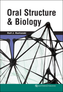
Oral Structure & Biology

Ralf J. Radlanski

Knowledge of the structures of the orofacial region from the macroscopic scale to the molecular level and pathologic changes to those structures enables practitioners to successfully treat patients or seek treatment options. This book presents the structural biologic foundations underpinning dental and oral medicine. Beginning with an overview of the anatomy of the mouth and moving on to the evolution of the oral structures and pre- and postnatal development of the oral cavity, related facial structures, and the teeth, this book describes each part of the orofacial region in terms of its morphology, tissue structure, cellular properties, and development. Functioning as both a textbook for dental students and a reference manual for experienced clinicians, this compendium of the structural biologic foundations of clinical work in dental and oral medicine allows practitioners to integrate current research in molecular biology into a solid framework of knowledge.

472 pp (softcover); 245 illus; ©2018; ISBN 978-0-86715-746-8 (B7468); $168


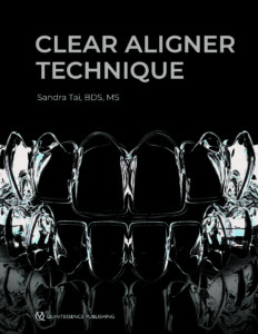
Clear Aligner Technique

Sandra Tai

Clear aligners are the future of orthodontics, but digital orthodontics evolves so rapidly that it is hard to keep pace. This book approaches clear aligner treatment from a diagnosis and treatment-planning perspective, discussing time-tested orthodontic principles like biomechanics and anchorage and demonstrating how to apply them to orthodontic cases using these appliances. Each chapter explains how to use clear aligners to treat a given malocclusion and teaches clinicians how to program a suitable treatment plan using available software, how to design the digital tooth movements to match the treatment goals, and finally how execute the treatment clinically and finish the case well. This clinical handbook will prepare orthodontists and dental students to exceed patient expectations with the most esthetic orthodontic appliance currently available.

320 pp; 1,344 illus; ©2018; ISBN 978-0-86715-777-2 (B7772); $218

Read more about Clear Aligner Technique here!

Clear Aligner Technique: Bringing Orthodontic Treatment into the Digital Era


Current Issues in Journals


prd_banner

Featured article: Immediate Postextraction Screw-Retained Partial and Full-Arch Rehabilitation: A 3-Year Follow-up Retrospective Clinical Study
Roberto Villa, Gabriele Villa, and Massimo Del Fabbro

A Histologic and Histomorphometric Retrospective Analysis of the Outcomes of Ridge Preservation Using Anorganic Bovine Bone Minerals and a Nonresorbable Membrane
Seiko Min, Marcelo Freire, Neema Bakshallian, Ivy Wu, and Homayoun H. Zadeh

Three-Year Results of a Randomized Controlled Clinical Trial Using Submucosally Veneered and Unveneered Zirconia Abutments Supporting All-Ceramic Single-Implant Crowns
Barbara Eisner, Nadja Naenni, Jürg Hüsler, Christoph Hämmerle, Daniel Thoma, and Irena Sailer

ijp_banner

Implant Survival in the Edentulous Jaw—30 Years of Experience. Part I: A Retro-Prospective Multivariate Regression Analysis of Overall Implant Failure in 4,585 Consecutively Treated Arches
Torsten Jemt

Passivity of Fit of a Novel Prefabricated Implant-Supported Mandibular Full-Arch Reconstruction: A Comparative In Vitro Study
Matthias Karl, Roberto Carretta, and Kenji W. Higuchi

Clinical Fit of Monolithic Zirconia Single Crowns
Stefanie Anke Rau, Michael Raedel, Aikaterini Mikeli, Martina Raedel, and Michael H. Walter

jomi_banner

THEMATIC ABSTRACT REVIEW: Use of Oral Implants for Cancer Reconstruction: The Role of the Approach to Vascular Flaps in Implant Outcomes
Martin Osswald

A Novel Experimental Dental Implant Permits Quantitative Grading of Surface-Property Effects on Osseointegration
Ernst B. Hunziker, Michael Spiegl-Habegger, Stefanie Rudolf, Yuelian Liu, Zhiyuan Gu, Kurt Lippuner, Nahoko Shintani, and Lukas Enggist

Systematic Review and Meta-Analysis of Hard Tissue Outcomes of Alveolar Ridge Preservation
Seyed Hossein Bassir, Muhanad Alhareky, Buddhathida Wangsrimongkol, Yinan Jia, MS, and Nadeem Karimbux

banner_ofph

Editorial: A Parting Perspective of the Journal’s Evolution
Barry J. Sessle

Peripheral Glial Cell Line–Derived Neurotrophic Factor Facilitates the Functional Recovery of Mechanical Nociception Following Inferior Alveolar Nerve Transection in Rats
Masahiro Watanabe, Masamichi Shinoda, Dulguun Batbold, Naoyuki Sugano, Shuichi Sato, and Koichi Iwata

Analgesic Effects of Intranasal Ketamine in Rat Models of Facial Pain
Rafaela Claudino, Carina Nones, Erika Araya, and Juliana Chichorro

Short clinical screening procedure for initial diagnosis of temporomandibular disorders
Georg Meyer

Multidisciplinary treatment-increase of vertical dimension combined with Invisalign treatment
Beatriz Solano Mendoza, Lorena Gómez García, Hourieh Pourhamid, and Enrique Solano

Correcting severe deep bite with the Invisalign appliance
Bärbl Reistenhofer, Fanny Triessnig, and Katharina Besser


Dental Meetings Quintessence Will Attend in October


AAOMS 100th Annual Meeting: Booth #414
hosted by the American Association of Oral and Maxillofacial Surgeons, October 11–13 in Chicago, Illinois

The USC International Restorative Dentistry Symposium
hosted by the Herman Ostrow School of Dentistry of USC, October 12–13 in Los Angeles, California

ADA 2018: Booth #704
hosted by the American Dental Association, October 18–22 in Honolulu, Hawaii

SCAD 2018 Annual Conference
hosted by the Society for Color and Appearance in Dentistry, October 19–20 in Newport Beach, California

AAMP 65th Annual Meeting
hosted by the American Academy of Maxillofacial Prosthetics, October 26–30 in Baltimore, Maryland

AAP 2018 Annual Meeting: Booth #1506
hosted by the American Academy of Periodontology, October 27–30 in Vancouver, Canada

ACP 2018 Annual Session: Booth #1506
hosted by the American College of Prosthodontists, October 31–November 3 in Baltimore, Maryland


Upcoming Quintessence Events


Posted in Announcement, Books, Journals, Multimedia, Promotions, Roundup, Special Offer, What's New | Tagged , | Leave a comment

3D Cephalometry: Is It Right for Your Orthodontic Practice?

Reading time: 15 minutes

As the body of research supporting the use of 3D radiography in orthodontics grows and the price of 3D CBCT technology wanes, many orthodontists find themselves asking an important question: Is 3D cephalometry right for my practice? Drs Katherine Kula and Ahmed Ghoneima, editors of the new book Cephalometry in Orthodontics: 2D and 3D, break down the decision-making process that practice owners should use to determine whether to upgrade to the third dimension.

3D imaging produces the most accurate representation possible, one that most closely matches our patients’ anatomy and the anatomical truth.

Ahmed Ghoneima, BDS, PhD, MSD
“Clinical orthodontics is currently transitioning from the 2D world to the 3D world,” explains Dr Kula, MS, DMD, MS. “As we evaluate the usefulness of 3D radiography in orthodontics, it’s important to consider that 3D cephalometry is only one of the many clinical advantages of 3D radiography. While 2D radiography provides an image for 2D cephalometry only, 3D radiography provides an image that can be used for either 3D or 2D cephalometry. 3D CBCT also provides digital information that allows for assessment of the position of teeth and various pathologies; the condition of the temporomandibular joint; and the volume and shape of the airways, alveolar bone, and soft tissue.”

“3D imaging significantly improves the diagnostic value of imaging in orthodontics,” adds Dr Ghoneima, BDS, PhD, MSD. “Advances in 3D imaging software programs have improved our ability to determine different treatment options, monitor changes over time, measure treatment outcomes more accurately, and predict and display the final treatment results. 3D imaging provides clear images with easily identifiable anatomical landmarks. It produces the most accurate representation possible, one that most closely matches our patients’ anatomy and the anatomical truth.”

When 3D CBCT technology first hit the market, it came with a price tag that made it largely inaccessible to most practitioners and their patients. However, recent innovations to the technology have focused on minimizing the impact of initial drawbacks such as the price of the machines and the amount of radiation used. These improvements make now the perfect time for orthodontists to begin considering adding this technology to their practice or upgrading to a dual-capability machine with both 2D and 3D sensors.

“Practitioners who already have radiography equipment in their practice should consider if the equipment is aging and may need replacement in the relatively near future,” Dr Kula advises. “In addition to speaking with company representatives, they should talk to other clinicians who currently have 3D CBCT machines in their practices. Even if your office’s current 2D radiographic equipment still has considerable life, it doesn’t hurt to start asking these questions now so you’ll be prepared in the future. It is also important to consider that for the new orthodontist who is just starting a practice and is particularly conscious of cost, a financial investment in 2D-only radiographic equipment makes it especially onerous to add or replace it with 3D-capable equipment in the near future.”

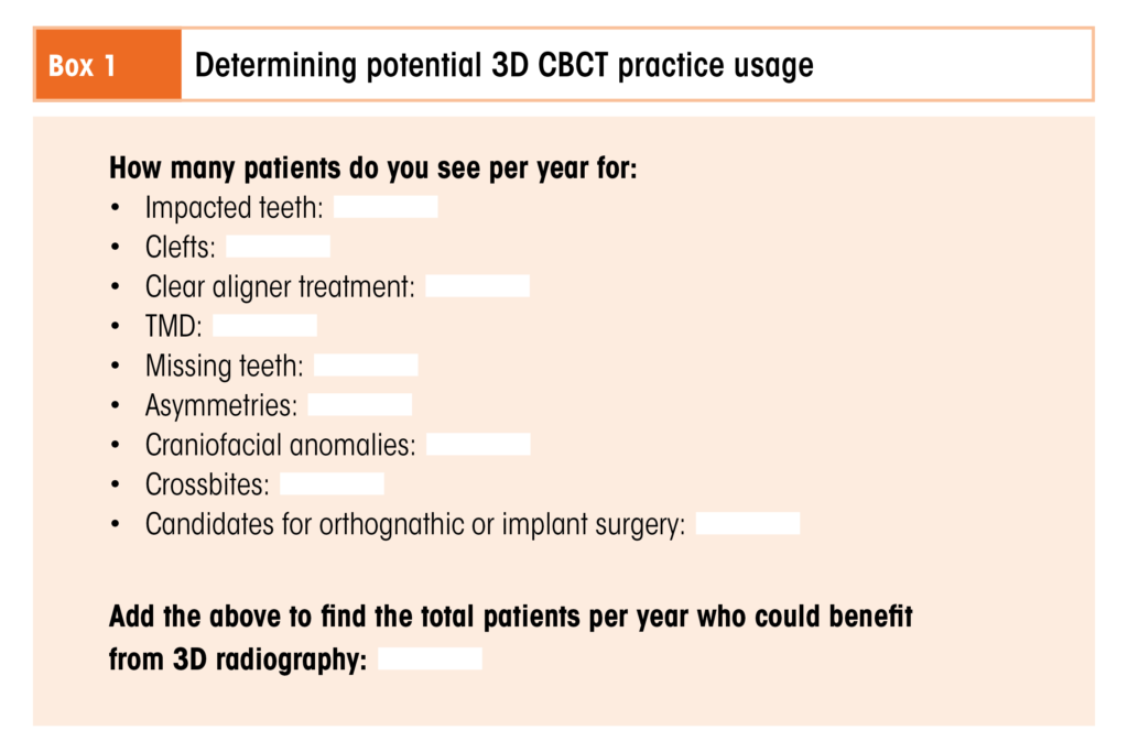
So how do clinicians decide whether to expand their practice’s capabilities to include 3D radiology? The list of questions curious clinicians should ask themselves is long: How many patients do you see who could benefit from having CBCT scans taken? How close is your current 2D radiographic equipment to needing replacement? How much does the unit you want cost, and how affordable will that cost be when it trickles down to your patients? Are there other local options for imaging to which you could refer your patients? The next sections will help you answer these questions and determine how you can incorporate 3D radiography into your practice.

Usage

The first step in deciding whether you should purchase a 3D CBCT machine for your orthodontic practice is to determine how often you would use the machine. But what are specific clinical indications where 3D radiography is preferred over 2D?

“There are several indications common to the orthodontic practice where 3D CBCT should replace 2D radiographs as part of the initial diagnostic records collection or as soon as possible after a problem is suspected,” Dr Kula explains. “These indications include suspected dental agenesis or impactions, missing teeth, various temporomandibular disorder signs or symptoms, facial and dental asymmetries, craniofacial anomalies, and crossbites. 3D CBCTs are also indicated for patients who will potentially have orthognathic surgery or implant placement.”

“Patients with airway disorders and obstructive sleep apnea should also receive a 3D radiograph,” Dr Ghoneima adds.

This compound odontoma (arrows) was discovered in an 11-year-old boy who was being evaluated for orthodontic treatment. (a) Axial view. (b) Cross-sectional views.

A 43-year-old man with fibrous dysplasia of the maxilla (arrows). The patient stated that his jaw began enlarging at 15 years of age. (a) Axial section of a CBCT scan through the maxilla. (b) Coronal view.

Eric Dellinger, DDS, MSD, breaks down his clinic’s 3D CBCT usage numbers in a chapter on cost comparison between 2D and 3D radiology in Cephalometry in Orthodontics: 2D and 3D: “The ability to refine diagnosis for patients with clefts, impacted canines, or craniofacial dysostosis might be a rarity in some offices. Within my own office, each year we see about 12 patients with clefts, 100 patients with impacted teeth, and 24 patients presenting for clear aligner treatment. Thus, we see only 136 patients per year who can potentially benefit from a 3D scan.”

Clinicians must consult their own numbers, as statistics from public data may be too general to be applicable. “Most research indicating the frequency of the types of patient anomalies that are indications for a 3D CBCT are usually based on the general population,” Dr Kula explains, “whereas these problems will naturally present more often in an orthodontic office than in the general population.”

Cost

Once you’ve established the prospective amount of usage for your clinic, you can now perform a cost analysis to see just how feasible the purchase of a 3D CBCT machine is for your practice. There are two important factors to consider when thinking about the cost of a 3D CBCT machine: the first is the cost of the machine itself, and the second is the cost of a CBCT scan to your patients.

“In my review of costs, I found a $44,000 increase in cost for a 3D machine relative to a traditional panoramic-cephalometric machine,” Dr Dellinger states. “This is significantly reduced from the difference of $100,000 or more that was the norm a few years ago. Nonetheless, this cost is a twofold increase in the normal cost for digital x-ray equipment, and thus the expense must be covered by supplemental treatment fees.”

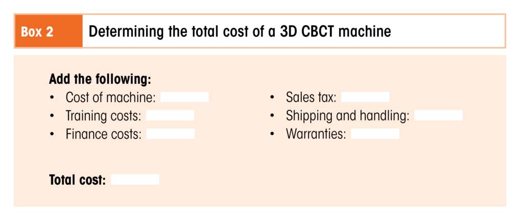
In addition to the sticker price of the machine, prospective buyers must also factor in additional costs like 3D software, warranties, and shipping and handling, as well as any applicable finance costs in order to calculate the full cost of implementing the technology into their practice. Staff members must receive training on the use of the machine, and the clinician or designated staff must also be trained on how to read and interpret the scans produced.

“With all of these extra charges,” Dr Dellinger says, “the final cost of the machine amortized over a 3-year period adds up to $2,000 for training (8 hours for 10 people) + $85,000 machine cost + finance costs of $6,711 (if the total of $85,000 is financed at 5% over 3 years) + $5,950 in sales tax (7% in Indiana) + $2,300 for shipping + $5,000 for warranties = $106,961. Thus, much like an automobile purchase, the total cost of the machine can be deceptively higher than the reduced sticker price may appear. As always, when approaching a significant expense for your practice, professional advice from your accountant may be very helpful in avoiding unpleasant surprises.”

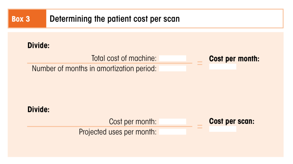
To determine the cost to your patients per scan, you can divide the total cost of the machine by the projected usage.

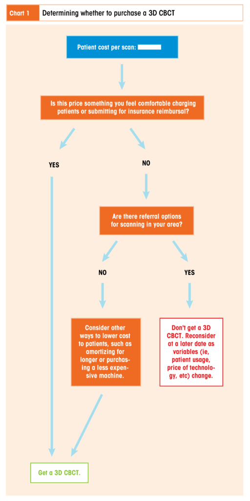
“If we assume a $100,000 cost divided over a 3-year amortization period,” Dr Dellinger calculates, “this comes to $2,778 per month. Assuming 20 uses per month, this leaves a cost of $138 per usage of the machine. By the same token, a 2D panoramic-cephalometric machine used 40 times per month at an initial cost of $45,000 (including some financing and training fees as well) comes to a price of only $62.50 per usage. Allowing these rough estimates, we see that the cost of 3D digital radiology will be about twice the cost of panoramic radiology. The choice of charging more for 3D scans and submitting this charge to medical insurance is also an option to defray costs. While the advantages of surgical evaluation of sinus structure and bone support can be invaluable in situations of cleft repair and impacted canines and third molars, and although this information is useful to orthodontists, the cost may exceed that of a referral to a radiology clinic for an MRI or 3D scan for evaluation of the patient, particularly in offices with fewer patients.”

Once you’ve done the numbers, one question remains: Is it worth it?

“Despite the additional monetary costs involved with 3D imaging, I find the personal reassurance of knowing the exact location of impactions and clefts to be of significant value,” Dr Dellinger emphasizes. “The confidence of knowing the precise positions of anatomy can lead to a much happier and confident clinician as well as a better treatment result for our patients. The cost of a 3D scan can thus produce several psychologic benefits, and these benefits can easily justify the additional cost for many clinicians. Given the advantages of 3D technology, it seems likely that we will continue to see improvements in this technology as well as increases in its use within dentistry and specifically within the practice of orthodontics.”

The Importance of Knowledge

Regardless of whether you decide to purchase a 3D CBCT machine for your own practice, it remains that we cannot go backward—only forward. We cannot ignore that there is now an option that can provide better, necessary information in many treatment circumstances. Clinicians must therefore prepare themselves to access and utilize this information through whatever means possible, whether by procuring their own 3D radiologic equipment or by referring their patients out for scans. Part of that preparation involves learning how to interpret the data produced in a 3D image using cephalometric principles.

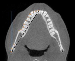
A 3D cone beam computed tomography (CBCT) section illustrating the differences in lengths if a 3D object is measured from one posterior lateral landmark to another anterior midline landmark from a 2D lateral perspective (blue line) or a 2D axial CBCT perspective (dashed black line). The actual length would be different if the perimeter of the object was measured (series of orange dots) because of the curve (3D CBCT perspective).

“Although not all clinicians will decide to include a 3D CBCT machine in their practice,” Dr Kula acknowledges, “they should be aware of the benefits that 3D radiology can provide to both the clinician and the patient. 3D radiology helps to optimize treatment outcomes and minimize risks such as the eruption of impacted teeth causing resorbed roots. Using 3D radiography, the clinician can ensure that patients are informed of the difficulty of their case and the reason for extra costs such as the extraction of impacted teeth. Clinicians who do not have a 3D CBCT machine in their office should be prepared to refer those patients to a clinic or imaging center that can provide a scan for pathology and a reading of the radiograph along with the image. In addition, the clinician should understand that 3D cephalometry is approximately a 1:1 image with minor magnification and that measurements from a 3D perspective are not always the same in a 2D measure, eg, profile or frontal.”

“Many clinicians,” Dr Ghoneima cautions, “may change their original treatment plan once they look at 3D images of their patients. This is because of hidden findings that could not be identified using conventional 2D images. For example, a patient may have significant root resorption due to ectopically impacted canines or a root fracture due to trauma. It is recommended that clinicians take a 3D radiograph if there is any uncertainty at all toward specific abnormal structures.”

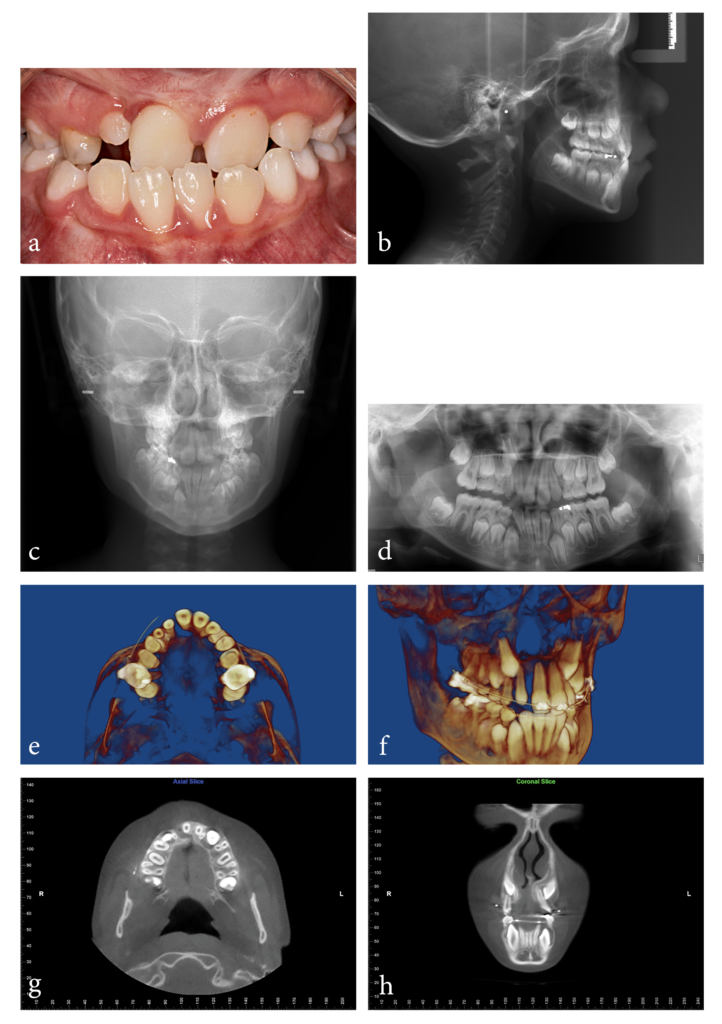
(a) Pretreatment intraoral photograph of a 9-year-old boy who was referred from the Cleft and Craniofacial Team with a diagnosis of cleft lip and no other significant medical history. Upon observing the thick frenum and a microdont maxillary lateral incisor, Dr Kula began to suspect there was an additional underlying issue. (b) Pretreatment lateral cephalogram. (c) Pretreatment frontal cephalogram. (d) Pretreatment panoramic radiograph. The panoramic radiograph showed probable impaction of the maxillary right canine and an indentation of the alveolar bone in the area between the microdont right lateral incisor and the right central incisor. The indentation appeared to continue toward the alveolar crest. At this point, the decision was made to take a 3D CBCT scan based on the appearance of the anterior maxillary arch, the frenum, nasal asymmetry, and the grooving observed in the panoramic radiograph. (e and f) Progress 3D CBCT reconstructions showing obvious clefting between the right central and lateral incisors from an axial view and a semilateral view. (g) Axial CBCT section showing the cleft with lack of alveolar continuity between the right lateral and central incisors. (h) Coronal CBCT section showing the position of the maxillary canines relative to the lateral incisor roots. The cleft was masked in the panoramic radiograph by two overlapping pieces of alveolar bone. The alveolar bone anterior to the maxillary and mandibular incisors was minimal. The alveolar bone palatal to the maxillary right central incisor was also minimal.

Information like that—information that can significantly influence a treatment plan—is invaluable to both the clinician and the patient. But in order to access it in full, clinicians must learn 3D cephalometric principles.

“The learning curve for 3D cephalometry is longer than for 2D cephalometry,” Dr Kula says. “For example, identification of a landmark from three perspectives—axial, coronal, and sagittal—takes a longer time. Landmark definitions are different because of the difference in perspective, and, unlike 2D cephalometry where landmark definitions are well established, landmark standards for 3D cephalometry still need to be developed and agreed upon. The learning curve for 3D cephalometry is shorter for clinicians who are introduced to it during residency programs compared with later, when practice management becomes a crucial focus for the orthodontist.”

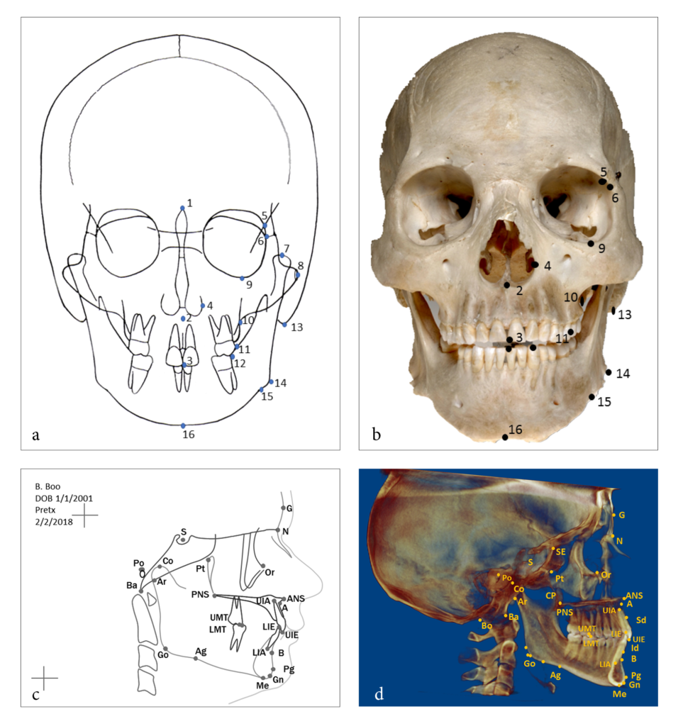
Note the differences between landmark locations on a 2D and 3D view and the addition of landmarks only visible using 3D CBCT. (a) Frontal landmarks on a manual tracing from a radiograph and (b) on a dry skull (only external landmarks can be marked). (c) 2D cephalometric tracing identifying (dots) and labeling (acronyms) landmarks. Labeling usually is not needed when the clinician is familiar with the landmarks. (d) Identification of landmarks on a 2D rendition of 3D CBCT.
There are several differences between the landmark locations on the 2D tracing compared with the 2D rendition of a 3D CBCT. One example is the gonion (Go), which is defined as the external angle of the mandible and located on the lateral radiograph by bisecting the angle formed by tangents to the posterior border of the ramus and the inferior border of the mandible (a line from menton to the posteroinferior border of the mandible).

“This learning curve gets easier to manage,” Dr Ghoneima adds, “once we have familiarized ourselves with this technology and how to manipulate the images to find the region of interest (ROI) in just a few clicks.”

It makes sense that as our technology changes, so must our knowledge base. Clinicians just starting to implement this technology must be aware that while 2D cephalometric principles may form the foundation of 3D cephalometry, the translation of knowledge is not one to one, and there is a great deal of new information to learn. Individual landmark definitions differ between 2D and 3D cephalometry, and there are important anatomical structures that can only be visualized using 3D imaging, leading to additional landmarks. And because landmark identification errors are considered a major source of cephalometric error, clinicians must obtain knowledge in 3D cephalometry that goes beyond understanding how to take a 3D CBCT scan.

The Future of 3D Cephalometry in Orthodontics

So will 2D radiology in orthodontics ever be completely replaced by 3D radiology, or will both continue to have unique benefits?

“Trying to answer that,” Dr Kula says, “reminds me of cartoons. The Flintstones makes me consider the wheel and how long it has been used. Despite numerous technological advances, the basic concept of the wheel still transports most of us. Several of the futuristic appliances from The Jetsons such as flat-screen televisions, video chatting, digital newspapers, and robotic vacuum cleaners have reached the common person, although others like flying cars, houses in the sky, and space tourism have not. It’s hard to predict what changes the near future will bring. But the answer of whether 3D radiology will replace 2D probably lies in patient need, ease of use, cost, radiation exposure, resolution, technological advancement, and marketing. As the technology improves, many of these variables are still changing, so only time will tell where it takes us.”

The space in time we occupy right now may feel closer to that of The Jetsons than The Flintstones, but every technological innovation we create is influenced by those that came before it. Likewise, 2D cephalometric knowledge forms the basis of 3D cephalometric knowledge. Cephalometry in Orthodontics: 2D and 3D creates a bridge between these two areas of knowledge so that clinicians can use both effectively, in turn enabling the clinician to practice 3D cephalometry no matter how they access the radiologic equipment.


Katherine Kula, MS, DMD, MS, retired as Jarabak Endowed Professor and Chairperson for the Department of Orthodontics and Oral Facial Genetics at the Indiana University School of Dentistry in Indianapolis, Indiana. A board-certified orthodontist, Dr Kula taught and practiced for more than 40 years. She has authored more than 100 papers and 18 chapters, edited 4 books and monographs, and is the recipient of several national research awards and grants. In addition to her activities in professional societies, Dr Kula organized the first meeting involving the American Dental Association (ADA) and the US Food and Drug Administration regarding the approval of dental therapeutics and devices, and her research is cited in the ADA’s stance concerning the effect of acidulated phosphate fluorides on dental materials. Her research interests currently include three-dimensional imaging, technology, music, growth and development of the craniofacial complex, and forensic facial reconstruction.


Ahmed Ghoneima, BDS, PhD, MSD, is Associate Professor of Orthodontics and Graduate Program Director of the College of Dental Medicine at Mohammed Bin Rashid University of Medicine and Health Sciences in Dubai, United Arab Emirates. He received his dental degree and master of science from Al-Azhar University in Cairo, Egypt, and his PhD from the Department of Orthodontics and Oral Facial Genetics at the Indiana University School of Dentistry in Indianapolis, Indiana, where he became a full-time faculty member. His research program focuses on airway analysis, applications of three-dimensional imaging in orthodontics, haptic technology, and the use of virtual reality in dental education. Dr Ghoneima is the recipient of numerous prestigious awards, including the Michael Matlof Memorial Teaching Fellowship Award and the Orhan C. Tuncay Teaching Fellowship Award, both from the American Association of Orthodontists Foundation, as well as the Academy for Academic Leadership Award. He has previously chaired the Indiana Section of the American Association for Dental Research and currently holds the position of Chair Elect for the American Dental Education Association section on orthodontics. He also serves as a reviewer for several peer-reviewed orthodontic journals.


Cephalometry in Orthodontics: 2D and 3D

Edited by Katherine Kula and Ahmed Ghoneima

 Cephalometrics has been used for decades to diagnose orthodontic problems and evaluate treatment. However, the shift from 2D to 3D radiography has left some orthodontists unsure about how to use this method effectively. This book defines and depicts all cephalometric landmarks on a skull or spine in both 2D and 3D and then identifies them on radiographs. Each major cephalometric analysis is described in detail, and the linear or angular measures are shown pictorially for better understanding. Because many orthodontists pick specific measures from various cephalometric analyses to formulate their own analysis, these measures are organized relative to the skeletal or dental structure and then compared or contrasted relative to diagnosis, growth, and treatment. Cephalometric norms (eg, age, sex, ethnicity) are also discussed relative to treatment and esthetics. The final chapter shows the application of these measures to clinical cases to teach clinicians and students how to use them effectively. As radiology transitions from 2D to 3D, it is important to evaluate the efficacy and cost-effectiveness of each in diagnosis and treatment, and this book outlines all of the relevant concerns for daily practice.

208 pp; 338 illus; ©2018; ISBN 978-0-86715-762-8 (B7628); US $118

Posted in Books, Feature, Orthodontics | Tagged , , , , , , | 2 Comments

The Time for Short Is Now: Today’s Short Implants and Why You Should Give Them a Chance

Reading time: 15 minutes

Short implants are currently experiencing a huge wave of commercial and clinical interest, with large implant companies like Nobel Biocare, Straumann, Dentsply Sirona, Biohorizon, and Zimmer Biomet all introducing their own offerings in recent years. Defined as implants with a designed intrabony length (DIL) equal to or less than 8 mm, short implants once occupied the far fringe of the implant market. Their recent rise to the mainstream may give some clinicians the false impression that these lengths of implants are new and relatively untested, but in reality, science established the value of short implants much earlier than the market did. Dr Douglas Deporter, a clinician and researcher who has devoted more than 30 years to the clinical study of short and ultra-short implants and who is the editor of a book on the topic, reflects on why he has dedicated his career to short implants. He also explains why their surge in clinical prominence today is wholly deserved.

A Periodontist’s Path to Implant Dentistry

“My interest in dental implants started almost 40 years ago,” explains Douglas Deporter, DDS, Dip Perio, PhD. “In 1979, I was a newly appointed assistant professor at the University of Toronto (U of T). The head of prosthodontics at my faculty, Prof George A. Zarb, proposed a site visit to investigate firsthand the rumors that an orthopedic surgeon by the name of Per-Ingvar Brånemark and his colleagues in Göteborg, Sweden, had developed and were testing a pure titanium threaded dental implant for tooth replacement. I was asked to be a member of the site visit team, and hence began my involvement in the implant field. Prof Zarb later organized a replication trial of Brånemark implants at U of T called ‘The Toronto Study.’ The early findings from The Toronto Study were supportive enough by 1982 that Prof Zarb and the Brånemark camp organized the first meeting in North America dedicated to the Brånemark implant concept, called the Toronto Conference on Osseointegration in Clinical Dentistry.”

My colleagues and I were thought to be way off base in thinking that implants could be much shorter in length than the standard lengths.

Douglas Deporter, DDS, Dip Perio, PhD
The Toronto meeting was significant in that it turned the tide on the scientific community’s opinion toward dental implants. Just 4 years prior in 1978, the US National Institutes of Health (NIH) and the Harvard University School of Dental Medicine had cohosted a conference with the intent of forming a consensus on dental implants. “The 1978 conference all but shut down interest in dental implants, which were seen by those involved as ‘snake oil treatments,'” Dr Deporter explains. “At the time, none of the participants were aware of Prof Brånemark’s work. But the impact of the 1978 conference was completely overruled by the findings presented at the Toronto meeting and the enthusiasm they generated.”

The invitation-only Toronto meeting primarily included prosthodontists and oral surgeons. Interestingly enough, periodontists were originally excluded from implant research and treatment due to the early opinion that the quality and quantity of soft tissue around implants was irrelevant. However, Dr Deporter managed to attend the Toronto meeting, and the experience changed his career path as a periodontist.

“I attended the Toronto conference with several members of our faculty,” Dr Deporter recalls, “including Prof Robert M. Pilliar, a biomaterials scientist responsible for the development of a highly successful cementless hip implant prosthetic device. Bob was intrigued by the idea of titanium dental implants, but not those like the Brånemark implant, which was a smooth machine-surfaced threaded device. Instead, he suggested to us that the same surface technology from his hip implant, which had a sintered porous surface (SPS) topography, could be applicable to dental implants and may offer some unique advantages over the threaded screw concept.”

The 7-mm-long SPSI Dr Deporter and his colleagues designed (Endopore, Innova Life Sciences) with healing cap (arrow). The DIL was only 5 mm because there was a 2-mm machine-turned collar segment. This size implant had a 20-year absolute survival rate of 90% when used to retain mandibular complete overdentures.

The SPS implant that Dr Deporter and his colleagues designed and tested was fabricated from titanium alloy and had a 5-degree taper to make it similar in shape to natural tooth roots. Early animal experiments indicated that the team was on the right track: They found that the sintered surface provided a much stronger bone-to-implant interface than a threaded implant with a machine-turned surface. The University of Toronto obtained a patent for the implant and awarded a license to Innova LifeSciences to produce it. Innova subsequently applied for approval from the US Food and Drug Administration (FDA).

“As expected, the FDA expressed doubt about the short implant lengths being used,” Dr Deporter recalls. “They insisted that our clinical work be duplicated by a small group of American dentists on American patients. The American clinicians, however, wanted longer implant lengths available to use when bone height allowed them to be used. Frankly, they did not believe that our short implant could work. In order to fulfill their request, Innova produced a 12-mm-long version, 12 mm being the maximum length possible with the implant’s designed 5-degree taper angle. We tried out some of these longer ones at U of T as well and, along with the US investigators, concluded that the longer length was of no advantage and may even underperform compared with our standard shorter length.”

Early Resistance

While the Toronto meeting galvanized interest in dental implants, it did so only for one type of dental implant—the Brånemark threaded implant—and only at lengths between 10 and 20 mm. Few in the dental community were as adventurous and willing to stray from the Brånemark path as Dr Deporter and his colleagues were. Further obstructing the acceptance of short implants were problems arising from early study designs and the way their results were presented, which unfortunately became the foundation of opinion for most clinicians and researchers.

Radiograph of two 4 × 4–mm ultra-short moderately rough threaded implants after 1 year in function in the posterior maxilla. (Courtesy of Dr Pietro Felice, University of Bologna, Italy; restoration by Dr Michele Diazzi, Bologna, Italy.)

“My colleagues and I were thought from the outset to be way off base in thinking that implants could be much shorter in length than were then seen to be safe and predictable,” Dr Deporter explains. “General opinion at the time was that if Brånemark implants were available in lengths ≥ 20 mm, how could an SPS implant with a DIL of just 5 mm work equally well? The conclusion was introduced that short lengths were inappropriate for Brånemark-type implants following early applications by less experienced clinicians such as those participating in The Toronto Study. However, later research verified that short Brånemark-type implants could have high survival rates even in the posterior maxilla as long as appropriate surgical modifications were made to the placement techniques.”

Once short implants have been fully integrated in bone, they behave just like longer implants.

Franck Renouard, DDS
“The poor reputation of short implants is—for the most part—based on early implant literature using the original Brånemark implant: A commercially pure titanium threaded screw with a machine-turned (ie, minimally rough) surface finish [References: 1, 2, 3, and 4],” says Past President of the European Association of Osseointegration and short implant advocate Dr Franck Renouard. “In each of these reports, the authors stated in their abstracts and/or conclusions that failure of short (ie, ≤ 10 mm) implants was higher than that of longer ones. This was enough to establish a dogma that was not easily challenged. However, a closer reading of these articles reveals that although the failure rates with short implants were higher, the difference compared with longer implants was less significant than the authors implied. For example, in the report by Friberg et al examining 4,641 implants, the failure rate of short maxillary implants covering everything from single crowns to full-arch reconstructions in fully edentulous patients was only 7%. Looking only at the results for partially edentulous patients in this study, the failure rate of short implants plummeted to 1.3%. Moreover, the authors stated that most of the failures were early—once short implants had been fully integrated in bone, they behaved just like longer implants. In 1991, these clinicians were not only using cylindric, machined-surface implants; they were also following a standard and identical drilling protocol for both short and standard-length implants and regardless of the bone density encountered.

(a) Scanning electron microscope (SEM) of a particle-blasted, acid-treated moderately rough threaded implant surface (original magnification ×2000). (b) SEM of an SPSI surface (original magnification ×200). (c) SEM of an SPSI surface after pull-out from a healed implanted site in a rabbit femur. Extensive bone ingrowth can be seen at the sheared bone-to-implant interface surface (original magnification ×300).

The first implants developed and tested successfully by Brånemark were under 8 mm in length.

Franck Renouard, DDS
“The reluctance of clinicians to use short implants persists today,” Dr Renouard continues, “and derives largely from their reading of statistical assessments of implant failure while neglecting other considerations like the patient’s sex, the size of the patient’s mouth, the risk of complications with more complex procedures, and the feasibility of such procedures being undertaken by non-specialists in a private practice setting. A quick search on PubMed in December 2016 identified 5,400 articles mentioning short implants, but the majority of articles focused on more complex solutions, relegating short implants to the rank of emergency fallback solutions only. Consequently, the mindset persists that the longer the implant, the more successful it will be both in the short and long term. Advanced, costly, and technique-sensitive collateral procedures often need to be performed to be able to use standard-length implants, such as autogenous (and other) bone block grafting, vertical alveolar ridge augmentation, mandibular nerve repositioning, and open sinus grafting. Interestingly, however, the first implants developed and tested successfully by Brånemark in the 1960s were all under 8 mm in length, and some were even shorter than 5 mm.”

One of the most important tenets of scientific research is to compare ‘apples to apples’ when comparing two things. It’s why controls are an important part of study design. Clinicians in these early studies were applying short implants in less favorable conditions than standard-length implants, but they weren’t controlling the reported data to accommodate those differences. This resulted in misleading comparisons between the efficacy of short versus standard-length implants. Hindsight and modern research also allow for the observation that the less-than-ideal machine-turned surface of early implant designs could have had a far more negative impact on outcomes than implant length.

This radiograph shows the status of two ultra-short implants after 2 years in function in
the resorbed posterior mandible. Note the increase in bone density adjacent to and between the two implants. Crestal bone has crept over the implant shoulders toward the neck segment.

“In 2005,” Dr Renouard states, “Hermann et al analyzed a large number of failed implant procedures and reported that short implants had usually been used in sites with low bone volume and density, whereas longer implants were nearly always placed in denser bone. This observation challenges implant length as the cause of failure. For example, did an implant in the posterior maxilla fail because of its length, or was the result caused by low bone density and the clinician’s failure to appropriately modify osteotomy preparation?”

The misleading presentation of success rates with short implants can be partially excused by simply not knowing then what we know now about important factors of success for endosseous implants. It can also be attributed in part to a phenomenon affecting all areas of research.

“The adaptation of these findings to comply with the general consensus at the time (ie, that implants shorter than 10 mm fail more often despite contrary objective data) is termed confirmation bias,” Dr Renouard explains. “This type of bias is a very common cognitive behavior: Once a decision has been made or a ‘fact’ learned, the human brain will always look for data that corroborate the preconceived notion in question and dismiss data challenging these notions. It is an unfortunate but frequent occurrence in many scientific fields.”

Current Knowledge

With the problems present in early research on short implants, where does the current literature stand? Dr Deporter’s book Short and Ultra-Short Implants provides a valuable update.

Some short implant designs have proven to be a legitimate substitute for longer implants used in conjunction with a dedicated open sinus floor elevation and grafting procedure.

Douglas Deporter, DDS, Dip Perio, PhD
“The most common location for short implant use is currently the resorbed posterior mandible,” Dr Deporter says, “but they can also be appropriate in the resorbed anterior mandible. In a posterior maxilla with resorption or a pneumatized sinus, some short implant designs have proven to be a legitimate substitute for longer implants used in conjunction with a dedicated open sinus floor elevation and grafting procedure. Using short or ultra-short implants in this last situation greatly reduces the associated risks, patient morbidity and anxiety, and cost. One recent investigator actually concluded that there is little justification for open lateral window sinus grafting where 5 mm of subantral bone remains preoperatively because a short implant with or without minimal indirect sinus floor elevation (eg, using hand osteotomes) will often work equally well. Using short or ultra-short implants can also simplify treatment in sites with alveolar ridge undercuts that would otherwise lead to apical bone fenestrations with longer implants.”

For this patient, short implants were used to retain a mandibular overdenture. Four 7-mm-long Brånemark-type implants were allowed submerged healing and subsequently connected with a customized bar structure. (a) Intraoral view at the 15-year recall of a patient’s four 7-mm-long implants and connecting bar. The peri-implant soft tissues appear healthy. (b) Radiograph at the 15-year recall. Peri-implant bone levels appear stable.

While these may seem like specific indications, experienced clinicians will recognize that no implant size or length can feasibly be used as a one-size-fits-all solution. Short and ultra-short implants therefore represent a valuable treatment option for situations that would generally require invasive augmentative procedures in order to place standard-length implants. Short implants also present several advantages, including lower risk of neurovascular damage in the posterior mandible, lower risk of sinus damage or infection in the posterior maxilla, simpler and less invasive surgical procedures, shorter treatment times, higher patient acceptance, and lower treatment costs for the patient. Even so, many clinicians may still feel more comfortable placing standard-length implants in conjunction with adjunctive surgical procedures, even in situations where short implants have proven successful. In the book, Dr Deporter and his colleagues address some of the common concerns that prevent clinicians from using short implants.

No implant size or length can feasibly be used as a one-size-fits-all solution.

“Until fairly recently,” Dr Deporter explains, “crown-to-implant (C/I) ratio was a major concern with short implants. From the outset of contemporary implant dentistry, it was theoretically and empirically decided that the C/I ratio should not exceed 1:1. This ratio was based on the established minimum crown/root ratio for healthy natural teeth; however, evidence has shown that due to the difference in nature between natural teeth retention and osseointegrated implants, this rule need not be applied. The perceived danger was a high levering effect at the bone crest that may result in bone microfractures and resorption. Recent research, however, has found that increasing the C/I ratio does not correlate positively with crestal bone loss, though evidence does point to there being critical upper limits for the C/I ratio that differ with different implant designs.”

Another concern is the fact that short implants cannot be treated the same as standard-length implants. Rather, a successful outcome largely depends on the clinician’s ability to correctly modify surgical protocols when placing short implants.

“Clinical data has shown that outcomes could be significantly improved by modifying standard threaded implant osteotomy preparation techniques,” Dr Deporter explains. “Preoperative evaluation of bone quality and density using CBCT can help the surgeon determine which modifications to make, which may include modified approaches such as an undersized osteotomy or a stepped osteotomy.”

(a) This patient was first treated with vertical ridge augmentation followed by placement of standard-length threaded implants and fixed prostheses. These later failed with loss of the grafted bone. (b) The patient was re-treated using two 4 × 4.5–mm self-tapping moderately rough threaded implants in the right mandible and two 4 × 4–mm self-tapping moderately rough threaded implants in the left mandible (all Global D implants). After removing the failed implants seen in a and without further bone grafting at the patient’s request, the sites were allowed to heal for 4 months before the ultra-short implants were placed. This radiograph was taken 1 year after the retreatment was completed. (c) This radiograph shows the right-side restoration after 4 years in function. (d) This radiograph shows the left-side restoration after 4 years in function.

(a) Treatment for this patient began with a vertical bone augmentation procedure. This procedure was unsuccessful, so the plan was changed to use three ultra-short 4 × 4–mm self-tapping moderately rough threaded implants. (b) A definitive prosthesis on its master cast. Note the accuracy and the polishing of the metal substructure to discourage dental plaque accumulation. (Courtesy of Dr Fabio Colombelli, Milan, Italy.) (c) A radiograph of the definitive prosthesis in place. (d) The clinical design of the definitive prosthesis showing optimal access for oral hygiene.

(a) Two single SPSIs were used to replace the maxillary right first premolar and first molar. The premolar site has a 7 × 4.1–mm (6-mm DIL) implant, while a 5 × 5–mm (4-mm DIL) implant was chosen for the molar site to avoid the need for a dedicated indirect sinus floor elevation procedure. Both implants (OT-F3) have an incorporated platform-switch feature. (b) Single anatomically correct metal-ceramic crowns were used for the two implants. (Restorations provided by Dr Ester Canton, Toronto, Ontario.)

(a) An ultra-short 5 × 4.1–mm (4-mm DIL) SPSI (Endopore) was placed in 3 mm of subantral bone using bone-added osteotome sinus floor elevation. A major portion of the implant length sits in new bone that formed following the sinus grafting. (b) A CBCT scan of the implant after 6 months of submerged healing prior to reentry and restoration. The majority of the implant is surrounded by new bone at this time. (Restoration by Dr Jeffrey Reynolds, Toronto, Ontario.)

Changing Perspectives

For the periodontist who has dedicated his career to the research and development of short implants, the changes occurring today are better late than never.

“Progress in dentistry is generally slow when it comes to applying research data to routine clinical practice,” Dr Deporter explains, “but it feels good to see change happening even if it has been at a snail’s pace. I have always felt that short would triumph in the long term, particularly when colleagues in countries other than the US started replicating and expanding upon our findings with SPS implants. Unfortunately, however, many dental practitioners—particularly in North America—still look to industry for answers rather than seeking out published independent research data. Luckily, most implant manufacturers have now seen the light and begun to produce short implant models, some as short as 4 mm. The fact that so many companies now produce short implants is a clear indication that there is a perceived high demand for them.”

However, there is also the potential to move backward and lose the ground so painstakingly gained. “I can only hope,” Dr Deporter says, “that any manufacturer that provides short implant options will have sufficient clinical trial data beforehand to provide defined proper protocols and limitations specific to their offerings. Short implants can be successful, but only if protocols are strictly followed and if clinicians pursue adequate information before using them. My fear is that the progress made to date may be undermined by high failure rates from clinicians who fail to understand that shorter does not necessarily mean simpler.

“Did I ever waver in my belief that short was good?” he continues. “That’s easy to answer: No. As they say, the proof is in the pudding.”


Douglas Deporter, DDS, Dip Perio, PhD, has been a full-time member of the Faculty of Dentistry at the University of Toronto since 1976. He has been involved in clinical research in implant dentistry for over 30 years and has published extensively on short and ultra-short implants, particularly those with sintered porous-surfaced (SPS) topography. He was coinventor of the original SPS implant (Endopore), the patent for which was assigned to the University of Toronto. Dr Deporter has given more than 140 invited international presentations. He also maintains a part-time private practice limited to periodontics and implant dentistry in Toronto, Ontario.


Short and Ultra-Short Implants

Edited by Douglas Deporter

Research has shown that short implants are not only a viable option but oftentimes a superior one that carries fewer risks for the patient and dentist, especially in resorbed jaw sites. As clinical trials continue to underscore the safety and efficacy of short implants, more dentists are considering their use with real interest, and this book provides the information clinicians need to incorporate short implants into their own practice. The book reviews the clinical effectiveness of short implants and then describes treatment protocols for the various types of short implants and their placement in different areas of the mouth. Case presentations demonstrate the recommended techniques and showcase the results.

168 pp (softcover); 334 illus; ©2018; ISBN 978-0-86715-785-7 (B7857); US $85

Posted in Books, Feature, Implant Dentistry | Tagged , | Leave a comment

Quintessence Roundup: September

Reading time: 10 minutes

September Monthly Special


Decision Making in Oral and Maxillofacial Surgery

Edited by Daniel M. Laskin and A. Omar Abubaker

This book assembles the decision-making acumen and experience of 30 oral and maxillofacial surgery specialists who have been recognized for expertise they have developed over years of patient treatment. Drawing on this body of knowledge mediated by experience, the contributors have synthesized their standard decision-making processes into annotated diagnosis and treatment algorithms. Combining “at-a-glance” understanding with detailed and authoritative discussion of the salient facts and features of more than 90 pathologic entities, these treatment algorithms are especially valuable for residents, recent graduates and others treating patients who present with unfamiliar signs and symptoms or with therapeutic problems in the oral and maxillofacial region.

268 pp; ©2007; ISBN 978-0-86715-463-4 (B4634); $120 Special price! $19


New Titles in Books


Cephalometry in Orthodontics: 2D and 3D

Edited by Katherine Kula and Ahmed Ghoneima

Cephalometrics has been used for decades to diagnose orthodontic problems and evaluate treatment. However, the shift from 2D to 3D radiography has left some orthodontists unsure about how to use this method effectively. This book defines and depicts all cephalometric landmarks on a skull or spine in both 2D and 3D and then identifies them on radiographs. Each major cephalometric analysis is described in detail, and the linear or angular measures are shown pictorially for better understanding. Because many orthodontists pick specific measures from various cephalometric analyses to formulate their own analysis, these measures are organized relative to the skeletal or dental structure and then compared or contrasted relative to diagnosis, growth, and treatment. Cephalometric norms (eg, age, sex, ethnicity) are also discussed relative to treatment and esthetics. The final chapter shows the application of these measures to clinical cases to teach clinicians and students how to use them effectively. As radiology transitions from 2D to 3D, it is important to evaluate the efficacy and cost-effectiveness of each in diagnosis and treatment, and this book outlines all of the relevant concerns for daily practice.

208 pp; 338 illus;©2018; ISBN 978-0-86715-762-8 (B7628); Now available! $118


The Tongue

Edited by Andreas Filippi and Irène Hitz Lindenmüller

As the largest organ in the oral cavity, the tongue not only plays a primary role in masticatory and speech function—it is also a significant indicator of health, demonstrating signs of both oral pathologies and diseases that can affect the entire body. Because no health care provider gets the opportunity to examine a patient’s tongue as often as the dentist, it is essential for dentists to recognize when there may be a problem with the tongue and what the problem is. In addition to an overview of tongue anatomy and general diagnosis and treatment recommendations, this book contains an atlas of more than 50 specific diseases and health concerns that may present signs and symptoms in the tongue. Each is outlined in a quick-reference table describing etiology, prognosis, and more and is accompanied by photographs of different ways the condition can present. A true diagnostic aid, this guide will allow clinicians to identify and address any abnormality a patient’s tongue may exhibit.

216 pp; 591 illus; ©2019; ISBN 978-0-86715-776-5 (B7765); $148 Special preorder price! $118
Available November 2018


Clinician’s Handbook of Oral and Maxillofacial Surgery, Second Edition

Edited by Daniel M. Laskin and Eric R. Carlson

There are frequent situations in which oral and maxillofacial surgeons find themselves in need of an immediate answer to a clinical problem. However, this can involve a time-consuming search for the appropriate reference source. This book continues the format of the previous edition by providing a single place to quickly find information on a diverse range of clinical topics, including dentoalveolar surgery, maxillofacial trauma, craniofacial anomalies, and oral pathology. All of the previous chapters have been updated, and new chapters on implantology, cleft lip and palate, maxillofacial reconstruction, oral squamous cell carcinoma, and cosmetic surgery have been added. Moreover, increasing the size of the book has allowed for the inclusion of many summary charts, tables, clinical photographs, and radiographs, which was not possible in the previous version. As a result, this new edition provides expanded information in an improved format.

Although this book is designed as a quick reference source, familiarizing oneself with its content in advance will both add to the reader’s general knowledge base and improve the ability to find information quickly in urgent situations. Residents in oral and maxillofacial surgery should find its content particularly useful during their clinical training, and the concise organization of the material should also be helpful to them in retaining information when subsequently preparing for the American Board of Oral and Maxillofacial Surgery.

624 pp (softcover); 374 illus; ©2019; ISBN 978-0-86715-730-7 (B7307); $168 Special preorder price! $134
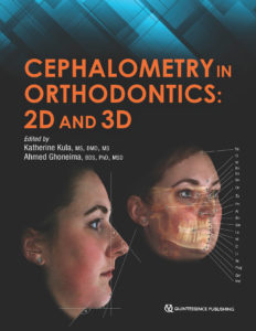
Available November 2018


Kratochvil’s Fundamentals of Removable Partial Dentures

Ting-Ling Chang, Daniela Orellana, and John Beumer III

In the 1960s, Professor F. J. Kratochvil recognized the importance of biomechanics in removable partial denture (RPD) design and used these principles to develop a new design philosophy. This “RPI system”—a clasp assembly consisting of a rest, a proximal plate, and an I-bar retainer—changed how clinicians approach partial denture design and is now used throughout the world. This textbook provides an overview of Kratochvil’s design philosophy and the basic principles of biomechanics it is based upon. Topics include components of RPDs and their functions, design sequences for maxillary and mandibular RPDs, and techniques for surveying and determining the most advantageous treatment position. A chapter dedicated to digital design and manufacturing of RPD frameworks highlights new technology in this emerging field. Additional topics include optimizing esthetic outcomes through attachments and rotational path RPDs as well as applying the RPI system to patients with maxillofacial defects. The authors provide illustrations of clinical cases throughout the book as well as an illustrated glossary of prosthodontic terminology. This textbook will prepare students and general practitioners to design and fabricate a biomechanically sound RPD framework for just about any dental configuration they encounter.

240 pp; 748 illus; ©2019; ISBN 978-0-86715-790-1 (B7901); $108 Special preorder price! $86
Available November 2018


Short and Ultra-Short Implants

Edited by Douglas Deporter

Research has shown that short implants are not only a viable option but oftentimes a superior one that carries fewer risks for the patient and dentist, especially in resorbed jaw sites. As clinical trials continue to underscore the safety and efficacy of short implants, more dentists are considering their use with real interest, and this book provides the information clinicians need to incorporate short implants into their own practice. The book reviews the clinical effectiveness of short implants and then describes treatment protocols for the various types of short implants and their placement in different areas of the mouth. Case presentations demonstrate the recommended techniques and showcase the results.

168 pp; 334 illus; ©2018; ISBN 978-0-86715-785-7 (B7857); $85 Special preorder price! $68
Available Summer 2018

Read more about Short and Ultra-Short Implants here!

http://www.quintpub.net/news/2018/08/the-time-for-short-is-now-todays-short-implants-and-why-you-should-give-them-a-chance/


Autologous Blood Concentrates

Arun K. Garg

Since the discovery of platelet-rich plasma (PRP) 25 years ago, interest in the use of autologous blood concentrates as adjuncts to surgical treatment has exploded. As more and more medically useful components of autologous blood concentrates have been identified, a host of unique acronyms such as PRF, CGF, PRGF, and more have surfaced, resulting in significant confusion among clinicians as to which material to use and when. Written by one of the original co-discoverers of PRP, this book tackles this issue of “too much information” by illuminating the science behind the clinical use of autologous blood concentrates as adjuncts to surgical treatment and helps to establish a foundation of practical knowledge for clinical use. The first part of the book summarizes the current literature from all aspects of medicine currently using autologous blood concentrates, showing both the possible applications as well as the limitations of these biologic materials. The second part of the book provides step-by-step instructions and richly illustrated treatment protocols for a number of applications for autologous blood concentrates specific to the practice of implantology and oral and maxillofacial surgery. Comprehensively researched and expertly written, this book is a must for clinicians who are just beginning to incorporate autologous blood concentrate treatment into their practice as well as experienced practitioners.

224 pp; 398 illus; ©2018; ISBN 978-0-99918-832-3 (B0007); US $199

Read more about Autologous Blood Concentrates here!

Autologous Blood Concentrates: Making Sense of all the Hype


Botulinum Toxin for Facial Harmony

Altamiro Flávio

The mastery of dentistry brings esthetic knowledge of the face that is applicable to more than just the teeth. In the process of performing a complete facial analysis, the practitioner can identify asymmetries and concerns localized to an area—such as the forehead, eyebrows, nose, or lower face—and offer Botox therapy to increase facial harmony. This book outlines the many clinical uses for Botox, with detailed illustrations and case presentations to support each procedure. The first part of the book covers systematic facial analysis, photographic documentation, and how to plan treatment. Special attention is paid to the anatomy and physiology of the face and the identification of injection points. Detailed treatment instructions for dosage, syringe type, and needle size are included for each procedure, as well as guidelines on how to evaluate results anthropometrically to determine whether esthetic treatment goals have been met. This stunning book will change the way you approach facial analysis and widen your esthetic treatment options for patients.

160 pp; 359 illus; ©2018; ISBN 978-0-86715-787-1 (B7871); Now available! $148

Read more about Botulinum Toxin for Facial Harmony here!

Botulinum Toxin for Facial Harmony: How this Unconventional Treatment Can Expand Your Practice


Oral Structure & Biology

Ralf J. Radlanski

Knowledge of the structures of the orofacial region from the macroscopic scale to the molecular level and pathologic changes to those structures enables practitioners to successfully treat patients or seek treatment options. This book presents the structural biologic foundations underpinning dental and oral medicine. Beginning with an overview of the anatomy of the mouth and moving on to the evolution of the oral structures and pre- and postnatal development of the oral cavity, related facial structures, and the teeth, this book describes each part of the orofacial region in terms of its morphology, tissue structure, cellular properties, and development. Functioning as both a textbook for dental students and a reference manual for experienced clinicians, this compendium of the structural biologic foundations of clinical work in dental and oral medicine allows practitioners to integrate current research in molecular biology into a solid framework of knowledge.

472 pp (softcover); 245 illus; ©2018; ISBN 978-0-86715-746-8 (B7468); Now available! $168


Clear Aligner Technique

Sandra Tai

Clear aligners are the future of orthodontics, but digital orthodontics evolves so rapidly that it is hard to keep pace. This book approaches clear aligner treatment from a diagnosis and treatment-planning perspective, discussing time-tested orthodontic principles like biomechanics and anchorage and demonstrating how to apply them to orthodontic cases using these appliances. Each chapter explains how to use clear aligners to treat a given malocclusion and teaches clinicians how to program a suitable treatment plan using available software, how to design the digital tooth movements to match the treatment goals, and finally how execute the treatment clinically and finish the case well. This clinical handbook will prepare orthodontists and dental students to exceed patient expectations with the most esthetic orthodontic appliance currently available.

320 pp; 1,344 illus; ©2018; ISBN 978-0-86715-777-2 (B7772); Now available! $218

Read more about Clear Aligner Technique here!

Clear Aligner Technique: Bringing Orthodontic Treatment into the Digital Era


Current Issues in Journals


prd_banner

Featured article: Immediate Postextraction Screw-Retained Partial and Full-Arch Rehabilitation: A 3-Year Follow-up Retrospective Clinical Study
Roberto Villa, Gabriele Villa, and Massimo Del Fabbro

A Histologic and Histomorphometric Retrospective Analysis of the Outcomes of Ridge Preservation Using Anorganic Bovine Bone Minerals and a Nonresorbable Membrane
Seiko Min, Marcelo Freire, Neema Bakshallian, Ivy Wu, and Homayoun H. Zadeh

Three-Year Results of a Randomized Controlled Clinical Trial Using Submucosally Veneered and Unveneered Zirconia Abutments Supporting All-Ceramic Single-Implant Crowns
Barbara Eisner, Nadja Naenni, Jürg Hüsler, Christoph Hämmerle, Daniel Thoma, and Irena Sailer

ijp_banner

Implant Survival in the Edentulous Jaw—30 Years of Experience. Part I: A Retro-Prospective Multivariate Regression Analysis of Overall Implant Failure in 4,585 Consecutively Treated Arches
Torsten Jemt

Passivity of Fit of a Novel Prefabricated Implant-Supported Mandibular Full-Arch Reconstruction: A Comparative In Vitro Study
Matthias Karl, Roberto Carretta, and Kenji W. Higuchi

Clinical Fit of Monolithic Zirconia Single Crowns
Stefanie Anke Rau, Michael Raedel, Aikaterini Mikeli, Martina Raedel, and Michael H. Walter

jomi_banner

Featured Article: Impact of Surface Chemistry Modifications on Speed and Strength of Osseointegration
Hyeon-Goo Kang, Yong-Soo Jeong, Yoon-Hyuk Huh, Chan-Jin Park, and Lee-Ra Cho

Thematic Abstract Review: Dental Implant Treatment of Atrophic Arches With or Without Augmentation
Jan-Eirik Ellingsen

Evaluation of Two 3D Printers for Guided Implant Surgery
Bruno Sommacal, Miodrag Savic, Andreas Filippi, Sebastian Kühl, and Florian M. Thieringer

banner_ofph

Editorial: A Parting Perspective of the Journal’s Evolution
Barry J. Sessle

Peripheral Glial Cell Line–Derived Neurotrophic Factor Facilitates the Functional Recovery of Mechanical Nociception Following Inferior Alveolar Nerve Transection in Rats
Masahiro Watanabe, Masamichi Shinoda, Dulguun Batbold, Naoyuki Sugano, Shuichi Sato, and Koichi Iwata

Analgesic Effects of Intranasal Ketamine in Rat Models of Facial Pain
Rafaela Claudino, Carina Nones, Erika Araya, and Juliana Chichorro

Short clinical screening procedure for initial diagnosis of temporomandibular disorders
Georg Meyer

Multidisciplinary treatment-increase of vertical dimension combined with Invisalign treatment
Beatriz Solano Mendoza, Lorena Gómez García, Hourieh Pourhamid, and Enrique Solano

Correcting severe deep bite with the Invisalign appliance
Bärbl Reistenhofer, Fanny Triessnig, and Katharina Besser


Dental Meetings Quintessence Will Attend in September


CDA North: Booth 1601
hosted by the California Dental Association, September 6–8 in San Francisco, California

Spear Annual Summit
hosted by Spear Education, September 13–15 in Scottsdale, Arizona

AO Mid-Atlantic Regional Meeting
hosted by the Academy of Osseointegration, September 14–15 in Gaylord, Maryland

AAID 67th Annual Conference: Booth #620
hosted by the American Academy of Implant Dentistry, September 26–29 in Dallas, Texas

ICOI World Congress XXXVI: Booth #310
hosted by the International Congress of Oral Implantologists, September 27–29 in Las Vegas, Nevada


Upcoming Quintessence Events


Posted in Promotions, Roundup, Special Offer, What's New | Leave a comment