Autologous Blood Concentrates: Making Sense of all the Hype

Reading time: 15 minutes

Since its discovery, platelet-rich plasma, or PRP, has developed into a $40-billion-a-year industry and has expanded from oral and maxillofacial surgery and implantology to other medical fields such as dermatology, orthopedic surgery, sports medicine, plastic surgery, and veterinary medicine. As the industry has grown, commercial interests and a lack of standardized language have led to a slew of similar and confusing acronyms: PRP, PRF, A-PRF, i-PRF, PVRG, PRFM, CGF, PC, hcPRP. . . How many of these have you heard of, and do you know the difference between them? Arun K. Garg, DMD, tells us how we got here.

In the Beginning There Was PRP

We expected healing to be a little better. What we found, though, is that healing was a lot better.

Arun K. Garg, DMD
“It started in the early 1990s when Robert E. Marx and I wanted to make bone sticky,” Dr Garg explains. “We needed to make it sticky in order to hold bone grafts together. Around that time they were selling something called fibrin glue in other countries, but it wasn’t available in the United States. So we decided to make our own autologous fibrin glue. We drew blood from the patient and spun the test tube in a centrifuge, forcing the plasma to the top, the erythrocytes to the bottom, and the fibrin to the middle. There were other blood components in the middle layer such as platelets and leukocytes, but we were mostly interested in the fibrin. When we applied this material to the bone, it held the bone graft particles together much better than without, and the process to make it only took minutes.”

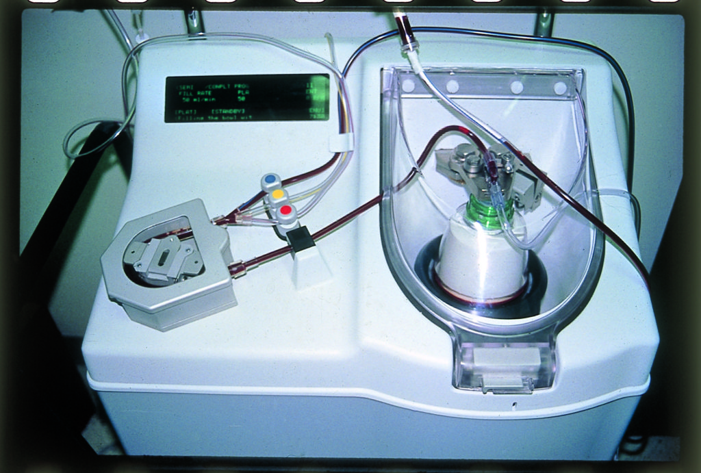
After that first success, they began implementing it more, using the equipment already present in the hospital operating room. “At that time, the heart pump we were using to centrifuge was about $80,000,” Dr Garg recalls, “but we didn’t have to buy it. It was already there in the hospital for cardiothoracic surgeons, so we used it. After we applied the material, the bone became sticky and much easier to handle.”

While the improvement to graft handling was immediately apparent, Dr Garg and his team were also eager to see whether this change would result in any improved healing. “We expected healing to be a little better because there would be less migration of the bone material,” he says. “What we found, though, is that healing was a lot better than expected. The healing time was almost twice as fast and the bone growth almost twice as dense. It was drastically better.”

Dr Garg emphasizes that to confirm results like this, he and Dr Marx would normally do several years of work and research before publishing. However, one of their residents at the University of Miami, Pairot Tayapongsak, published on it in 1994 and called the material autologous fibrin adhesive. (He did not use an acronym in this paper.)

“By referring to the material as autologous fibrin adhesive, Dr Tayapongsak put emphasis on the fibrin component of the material,” Dr Garg explains. “And the fibrin content did explain part of the improved healing by way of holding the bone material together, but it didn’t explain the whole result. What we discovered was that the effect wasn’t just due to the fibrin content: it was also influenced by the concentrated platelet content.”

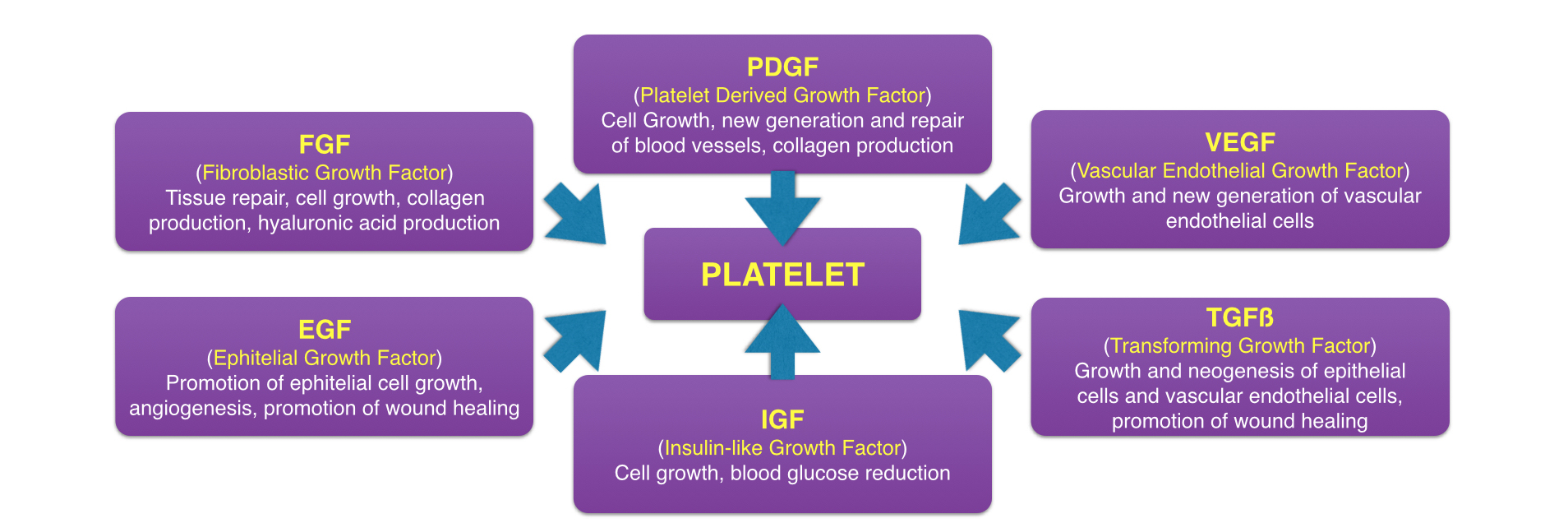
At that time, medical professionals were well aware of the fact that platelets control the first step of healing by being the first to arrive at the site of injury and inducing clotting. However, what was not well known at the time is that platelets also initiate the second step of healing—wound repair—by releasing growth factors at the area of injury. So when Drs Marx and Garg’s team increased the number of platelets to the surgical area by fivefold, they also inadvertently increased the number of platelet-derived growth factors by fivefold. By amplifying these growth factors, they effectively amplified wound healing at the surgical site, resulting in both increased speed and quality of healing.

The known types of growth factors released by platelets.

All of this brings us back to the name. “After this discovery,” Dr Garg said, “we decided that, while fibrin was important for its mechanical impact, it wasn’t the star of the show. So we switched gears to give star billing to the platelets by calling it platelet-rich plasma, or PRP.”

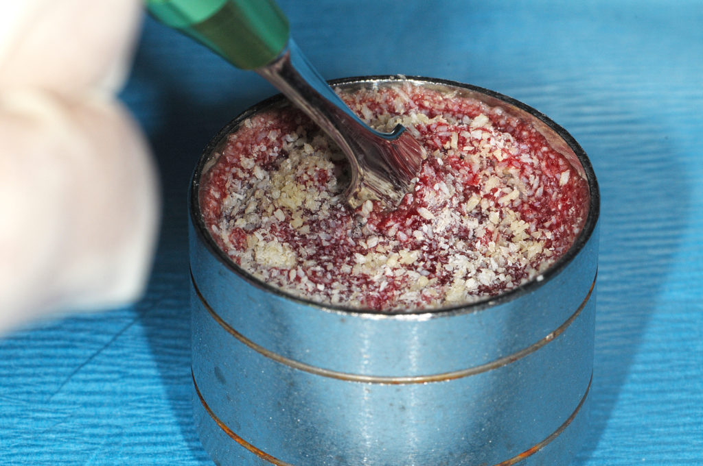
PRP exudate used to hydrate bone graft material.

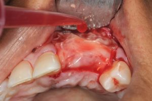
PRP exudate used to hydrate a membrane after placement.

Now that they understood why the PRP was having the effect it was having, Dr Garg and his team began experimenting with different surgical configurations of the material. The original configuration involved adding anticoagulant to the drawn and centrifuged blood to keep it from clotting before use and then inducing clotting once the material was added to the bone graft material. This became known as PRP liquid and can be used in long surgical procedures or as an injectable. The next configuration they experimented with was allowing the PRP to clot before use. They found that the clotted PRP was best applied during short surgeries because, if the material is left to clot for too long, too many of the growth factors are released prematurely and left behind in the receptacle when the material was removed and used.

“The clotted material can’t be used to hydrate the bone,” Dr Garg says, “but you can certainly add it into an extraction socket. This became known as PRP gel. You can also flatten the clotted PRP into a fibrinous membrane and use it to contain bone graft material, and this became known as a PRP membrane. We also published on using the gel in sinus lifts for bone formation: gel on one side, bone on the other. On the crestal side, it can be hard to get bone graft material into that small opening without tearing the sinus membrane. But with PRP gel, you can achieve the necessary stability, and it’s easier to pack it in. The gel will either push up on the sinus membrane or come back out of the osteotomy, but it doesn’t have enough ‘body’ to actually tear the sinus membrane. Before PRP gel, a crestal sinus lift was a very technique-sensitive procedure; however, now it’s ubiquitous.”

All of these were names for the same substance, just in different configurations for different applications, and its versatility as an autogenous material that could be used to enhance wound healing caused it to spread rapidly.

The Market Success and Spread of Autologous Blood Concentrates

After the success seen in oral surgical applications, the use of PRP quickly spread from dentistry to medicine, particularly in specialties like orthopedic surgery, sports medicine, and veterinary medicine. In sports medicine and orthopedic surgery, the use of PRP achieved high demand as an adjunctive treatment for sport-related injuries in professional athletes, as the decreased healing time was especially valuable in getting athletes off the bench and back onto the field. Similarly, PRP spread to veterinary medicine and was used in injured racehorses. The cost of PRP treatment was an easy choice for an athlete with a multimillion-dollar season contract or in a horse like recent Triple Crown winner Justify, who is worth an estimated $75 million. But in order for PRP to become the powerhouse of an industry it is today, the technology needed to be financially accessible for a majority of health providers, and an $80,000 price tag for a centrifuge was far too steep for in-office use.

The plasmapheresis machine originally used to separate PRP.

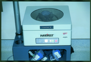
A Harvest Technologies in-office centrifuge system.

When Drs Marx and Garg first began using PRP, they were using equipment already available at the hospital where they were performing their surgeries, so there were no start-up costs for implementing the technology—just the operation costs of test tubes and paying a perfusionist to operate the centrifuge during the surgery. Once the use of PRP began to spread, a company called Harvest Technologies released an in-office centrifuge machine that cost just $8,000—a fraction of the price of the original centrifuge Drs Marx and Garg had started using in the hospital operating room. However, there was a special catch to their business model: in order to turn a profit while selling the centrifuge below market value, they cranked up the cost on the test tubes to $350 apiece. The amount spent on single-use test tubes adds up quickly, and, by holding the customer hostage by way of limiting which test tubes could be used in the machine, the company also ensures a longer tail of revenue from each customer. Harvest Technologies put its centrifuge machines in almost every major hospital in the country because they knew that once surgeons started using PRP, they would have to keep buying the test tubes. And it worked: in just 8 years, a Japanese group purchased Harvest Technologies—a company with a single product, the centrifuge and its test tubes—for $80 million.

“After the sale of Harvest Technologies,” Dr Garg explains, “everyone started thinking about how they’d like to make $80 million off one product, so the market exploded. In the US alone, there are probably 40 different manufacturers of these centrifuges now. We started seeing centrifuges for $4,000, $2,000, and $800 with test tubes for $200, $100, and $40 each. The prices were all over the place for the same technology. It was similar to the Keurig model: make the brewing machines inexpensive enough to become ubiquitous, then rely on the higher profit margin of the single-use coffee pods. And everyone says, ‘Our coffee tastes better.’ There have even been companies who try to sell $40 test tubes over $0.30 test tubes by saying—I kid you not—’my test tubes are made of Moroccan glass.’ Who cares if it’s made from Moroccan glass? It’s a test tube!”

So with all the flashy options available, which machine does the original co-discoverer of PRP use? “In our lectures,” Dr Garg says, “we demonstrate the machine by whichever company sponsors the course because the technology is all the same. But the one I use daily in my six practices is a $1,500 machine with generic $2 test tubes. If I were to use a machine with more expensive test tubes, that cost would get passed on to my patients. The cost of a $1,500 machine is nominal when spread out in a practice, so with my setup the cost of PRP treatment is just $2 for the test tube and the needle to draw the blood.”

What’s in a Name? The Many Terms for Autologous Blood Concentrates

As the market became saturated with centrifuge options, a new strategy to differentiate products and capture market share emerged: giving the material produced a different name.

“The first second-generation autologous blood concentrate was PRF, which stood for platelet-rich fibrin,” Dr Garg explains. “The term PRF was coined by a Moroccan-French doctor who also went on to coin other terms such as leukocyte-platelet–rich fibrin (L-PRF), advanced PRF (A-PRF), and injectable PRF (i-PRF), and later A-PRF+ and i-PRF+. A Spanish oral surgeon coined the term plasma rich in growth factors (PRGF), and in Italy and Korea they began calling the material concentrated growth factors (CGF). So now we have all of these terms in an industry that lacks standardization of nomenclature, and clinicians and patients alike are understandably confused.”

So is there a difference between all of these materials?

“The first and most obvious difference is whether or not you use an anticoagulant,” Dr Garg explains, “because that affects the clotting and centrifugation of the material. By instruction, PRF is described as never using an anticoagulant, and because of that much of the literature today states that the major difference between PRP and PRF is anticoagulant inclusion. However, when we first started using PRP, we often forewent the use of an anticoagulant—the decision was always based on the particular application and how quickly we were using the material. So to say PRP is always used with an anticoagulant would be completely inaccurate. We added the anticoagulant because we found that, for lengthy surgical procedures where the material may be drawn long before its use, if we allowed the material to coagulate for too long, too many of the platelet-derived growth factors would be released early in the receptacle rather than at the surgical site.”

“The other differences,” he continues, “relate to the centrifuge itself. By altering the rotations-per-minute (RPM) of the machine, you can affect the concentration. Also important, however, are the size of the rotator arm, how long you spin the material for, and the angle of the test tube in the centrifuge. One thing that you absolutely cannot do is take the production recommendations from one manufacturer’s centrifuge, apply it to another, and expect a good result, because manufacturers set their speed and length of time recommendations based on the specifications of that machine’s rotator arm and angle of the test tube.”

According to marketers, you can only make certain materials using their specific centrifuges. But according to Dr Garg, you can customize the centrifuge process to create any of these materials. “By modifying spin speeds and time, different components of the middle layer can be slightly increased or slightly decreased in concentration. What the clinician must do is identify which of the configurations of autologous blood concentrates will have the effect he or she desires in each individual case. For example, perhaps in one case soft tissue healing is your main concern and increased growth factors are your focus, so you create and apply a PRP gel membrane. In another case, you’re performing a ridge augmentation where the mechanical benefit of increased fibrin content would be clinically appreciated, so you create and apply a clotted PRP membrane. Each configuration has its set of preferred applications, and you have to be able to identify which configuration is needed in each case in order to achieve the best possible result.”

But with so much contradictory information out there, how do clinicians parse through the advertising narratives from manufacturers to find the instruction they need to use all of these different materials correctly? In an effort to redirect the conversation back to evidence-backed science, Dr Garg has written an entire book on just that subject.

Setting the Record Straight

It had become impossible for clinicians to separate the science from the sell.

Arun K. Garg, DMD
After they first discovered the benefits of PRP and pioneered the first clinical applications, Drs Marx and Garg spent the next decade giving CE courses at a rate of about one per week to train other clinicians on the use of PRP as well as publishing nearly a dozen peer-reviewed journal articles on the topic. In 2005, they gathered their research, clinical results, and teaching materials into the book Dental and Craniofacial Applications of Platelet-Rich Plasma (Quintessence).

“Once we had the book out there,” Dr Garg recalls, “we stopped doing the CE courses. After we stopped lecturing on it, a lot of misinformation came in to take our place, and that led to a lot of confusion. I felt it was time for an update. I wanted to explain things in a way that would minimize the confusion among clinicians and reduce the effect of ‘paralysis by analysis’—where you overanalyze something so much that you get overwhelmed and never actually use it. The other issue was that commercial interests had completely taken over the conversation. It had become impossible for clinicians to separate the science from the sell. I wanted to get the information back on track.”

Autologous Blood Concentrates (Garg Multimedia Group, 2018) is the result of those efforts. With this book, Dr Garg has stepped back in as a strong, objective voice capable of helping to guide the present conversation surrounding autologous blood concentrates. In the spirit of setting aside his own personal interests as co-discoverer of PRP, he has even attempted to forego the term PRP and instead adopted autologous blood concentrates as a generic term for the materials, while also humbly admitting how hard the habit was to break:

“In this book,” he explains, “I’ve tried to minimize the chest pounding and salesmanship that has degraded the conversation into debates on nomenclature and instead focus on the science, biology, and clinical applications. I wanted the book to reach clinicians regardless of which machine or nomenclature they were most familiar with, so I didn’t want whichever acronym I used to be a roadblock for anyone. That’s why the title of the book is Autologous Blood Concentrates. I’ve tried to use that terminology as often as possible in the book. However, because I’ve called it PRP for 25 years, there are several cases where I’ve slipped and called it PRP. The reader is welcome to simply substitute ‘autologous blood concentrate’ in these instances; the information remains the same.
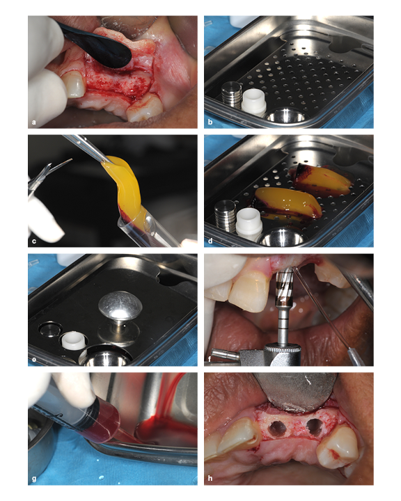
(a) The ridge is too thin for implant placement, so augmentation is required. (b) A PRP membrane kit. (c) PRP gel being removed from the test tube. (d and e) PRP gel placed in the membrane disk. (f) The ridge is widened by running the bur in reverse. (g) PRP exudate is extracted from the membrane dish using a syringe. (h) The prepared socket.
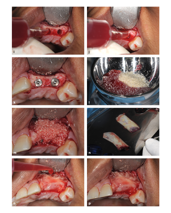
(i and j) The PRP exudate is used to hydrate the prepared socket. (k) Implant placement. (l) The freeze-dried bone allograft particles are hydrated with PRP exudate and mixed. (m) The ridge is packed with the bone graft particle and PRP exudate mixture. (n) The PRP membrane is uncovered in the membrane kit. (o) The PRP membrane is placed and then hydrated with PRP exudate. (p) The final view.

“This material has taken off,” he continues, “because it’s natural, it’s inexpensive, it’s safe, and it has so many applications. I wrote this book to make the information more accessible to clinicians, but I especially wanted to make the material itself accessible. While it must be remembered that it can’t make a bad surgery good, it will make a good surgery better, and that is what’s worthwhile.”

While it may not be made of Moroccan glass, Dr Garg’s book brings value to the field by redirecting the conversation of autologous blood concentrates out of the profit margins and back to the patient benefits. It represents a road map clinicians can use to navigate the plethora of available information on these materials and translate that information into clinical decision-making. Similarly, Dr Garg’s book provides a model for researchers on how these materials can be described and dealt with in the literature in order to minimize confusion as the field continues to evolve and press forward. In an industry that is still finding its footing with regard to communication, standardization, and replication of results, this book will certainly have a meaningful impact on the legacy of autologous blood concentrates.
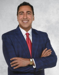

Arun K. Garg, DMD, is considered one of the world’s foremost authorities on bone biology, harvesting, and grafting for dental implant surgery. Alongside Dr Robert E. Marx, he pioneered the use of platelet-rich plasma (PRP). He received his dental degree from the University of Florida College of Dentistry in Gainesville, Florida, and completed his residency at the University of Miami Jackson Memorial Hospital in Miami, Florida. He went on to serve for almost two decades as Professor of Surgery in the Division of Oral and Maxillofacial Surgery and as Director of Residency Training at the University of Miami Leonard M. Miller School of Medicine, where he was frequently awarded faculty member of the year by his residents. Dr Garg has published 8 medical textbooks and over 150 journal articles is currently the president of the International Dental Implant Association. An international lecturer, he also maintains six private practices throughout southern Florida and is the founder of Implant Seminars, a leading dental continuing education company.


Autologous Blood Concentrates

Arun K. Garg

Since the discovery of platelet-rich plasma (PRP) 25 years ago, interest in the use of autologous blood concentrates as adjuncts to surgical treatment has exploded. As more and more medically useful components of autologous blood concentrates have been identified, a host of unique acronyms such as PRF, CGF, PRGF, and more have surfaced, resulting in significant confusion among clinicians as to which material to use and when. Written by one of the original co-discoverers of PRP, this book tackles this issue of “too much information” by illuminating the science behind the clinical use of autologous blood concentrates as adjuncts to surgical treatment and helps to establish a foundation of practical knowledge for clinical use. The first part of the book summarizes the current literature from all aspects of medicine currently using autologous blood concentrates, showing both the possible applications as well as the limitations of these biologic materials. The second part of the book provides step-by-step instructions and richly illustrated treatment protocols for a number of applications for autologous blood concentrates specific to the practice of implantology and oral and maxillofacial surgery. Comprehensively researched and expertly written, this book is a must for clinicians who are just beginning to incorporate autologous blood concentrate treatment into their practice as well as experienced practitioners.

224 pp; 398 illus; ©2018; ISBN 978-0-99918-832-3 (B0007); US $199


This article was written by Caitlin Davis, Quintessence Publishing.

©2018–2019 BY QUINTESSENCE PUBLISHING CO, INC. PRINTING OF THIS DOCUMENT IS RESTRICTED TO PERSONAL USE ONLY. NO PART MAY BE REPRODUCED OR TRANSMITTED IN ANY FORM WITHOUT WRITTEN PERMISSION FROM THE PUBLISHER.

Posted in Feature, Implant Dentistry, Multidisciplinary, Oral and Maxillofacial Surgery | Tagged , , , , , , , , , | 5 Comments

Quintessence Roundup: August

Reading time: 9 minutes

August Monthly Special


Critical Thinking: Understanding and Evaluating Dental Research, Second Edition

Donald Maxwell Brunette

The second edition of this classic textbook prepares readers to identify the information they need, read it from a critical vantage point, and assess the soundness of the conclusions. It introduces issues of logic, statistics, measurement, research design, and argument. Invaluable for anyone who wishes to become a discerning reader of dental literature.

324 pp (softcover); 90 illus; ©2007; ISBN 978-0-86715-426-9 (B4269); $48 Special price! $24


New Titles in Books


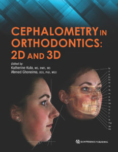
Cephalometry in Orthodontics: 2D and 3D

Edited by Katherine Kula and Ahmed Ghoneima

Cephalometrics has been used for decades to diagnose orthodontic problems and evaluate treatment. However, the shift from 2D to 3D radiography has left some orthodontists unsure about how to use this method effectively. This book defines and depicts all cephalometric landmarks on a skull or spine in both 2D and 3D and then identifies them on radiographs. Each major cephalometric analysis is described in detail, and the linear or angular measures are shown pictorially for better understanding. Because many orthodontists pick specific measures from various cephalometric analyses to formulate their own analysis, these measures are organized relative to the skeletal or dental structure and then compared or contrasted relative to diagnosis, growth, and treatment. Cephalometric norms (eg, age, sex, ethnicity) are also discussed relative to treatment and esthetics. The final chapter shows the application of these measures to clinical cases to teach clinicians and students how to use them effectively. As radiology transitions from 2D to 3D, it is important to evaluate the efficacy and cost-effectiveness of each in diagnosis and treatment, and this book outlines all of the relevant concerns for daily practice.

208 pp; 338 illus;©2018; ISBN 978-0-86715-762-8 (B7628); $118 Special preorder price! $94
Available Summer 2018


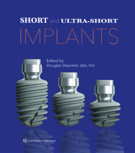
Short and Ultra-Short Implants

Edited by Douglas Deporter

Research has shown that short implants are not only a viable option but oftentimes a superior one that carries fewer risks for the patient and dentist, especially in resorbed jaw sites. As clinical trials continue to underscore the safety and efficacy of short implants, more dentists are considering their use with real interest, and this book provides the information clinicians need to incorporate short implants into their own practice. The book reviews the clinical effectiveness of short implants and then describes treatment protocols for the various types of short implants and their placement in different areas of the mouth. Case presentations demonstrate the recommended techniques and showcase the results.

168 pp; 334 illus; ©2018; ISBN 978-0-86715-785-7 (B7857); $85 Special preorder price! $68
Available Summer 2018


Autologous Blood Concentrates

Arun K. Garg

Since the discovery of platelet-rich plasma (PRP) 25 years ago, interest in the use of autologous blood concentrates as adjuncts to surgical treatment has exploded. As more and more medically useful components of autologous blood concentrates have been identified, a host of unique acronyms such as PRF, CGF, PRGF, and more have surfaced, resulting in significant confusion among clinicians as to which material to use and when. Written by one of the original co-discoverers of PRP, this book tackles this issue of “too much information” by illuminating the science behind the clinical use of autologous blood concentrates as adjuncts to surgical treatment and helps to establish a foundation of practical knowledge for clinical use. The first part of the book summarizes the current literature from all aspects of medicine currently using autologous blood concentrates, showing both the possible applications as well as the limitations of these biologic materials. The second part of the book provides step-by-step instructions and richly illustrated treatment protocols for a number of applications for autologous blood concentrates specific to the practice of implantology and oral and maxillofacial surgery. Comprehensively researched and expertly written, this book is a must for clinicians who are just beginning to incorporate autologous blood concentrate treatment into their practice as well as experienced practitioners.

224 pp; 398 illus; ©2018; ISBN 978-0-99918-832-3 (B0007); US $199

Read more about Autologous Blood Concentrates here!

http://www.quintpub.net/news/2018/07/autologous-blood-concentrates-making-sense-of-all-the-hype/


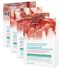
Decision Making for Retreatment of Failures in Dental Medicine (DVD-ROM)

Giorgio Tabanella (editor)

When patients present to the dental clinic for retreatment, there is little room for error. Cases requiring retreatment may have already resulted in complications, failures, and even iatrogenic damage. It is therefore in the best interest of patients and for the discipline that clinicians share protocols, techniques, and innovations used in retreatment to improve the quality of care and avoid further error. The clinical cases depicted in this DVD compendium all lay bare two things: first the medical mistake and then the innovative fix that allowed for a successful outcome at retreatment.

DVD compendium: 4 DVDs; Format: NTSC/PAL; ©2018; ISBN 978-1-78698-006-9 (C0975); US $230 for the four-volume set or $80 each


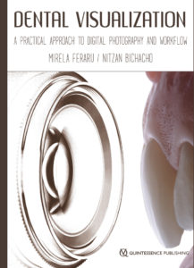
Dental Visualization: A Practical Approach to Digital Photography and Workflow

Mirela Feraru and Nitzan Bichacho

This book concentrates the authors’ extensive knowledge of clinical dental photography into a comprehensive, practical, and relevant guide for the practicing clinician. What sets this book apart from other titles available on dental photography is that it focuses not on artistic photography but rather on the concept of dental visualization: dental photographs that are taken and used as a clinically relevant tool. The major principles of dental photography are broken down into concise chapters and illustrated with high-quality, detailed images. Topics covered include photographic equipment, camera components, digital documentation, and troubleshooting. Simplified protocols for achieving high-end photographic results are presented step by step, and considerations for the different dental specialties are also described. This book promises to be an invaluable tool for both general practitioners and specialists to elevate their services provided by using dental photography.

248 pp; 556 illus; ©2018; ISBN 978-1-78698-004-5 (B9104); Now available! $158


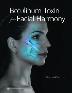
Botulinum Toxin for Facial Harmony

Altamiro Flávio

The mastery of dentistry brings esthetic knowledge of the face that is applicable to more than just the teeth. In the process of performing a complete facial analysis, the practitioner can identify asymmetries and concerns localized to an area—such as the forehead, eyebrows, nose, or lower face—and offer Botox therapy to increase facial harmony. This book outlines the many clinical uses for Botox, with detailed illustrations and case presentations to support each procedure. The first part of the book covers systematic facial analysis, photographic documentation, and how to plan treatment. Special attention is paid to the anatomy and physiology of the face and the identification of injection points. Detailed treatment instructions for dosage, syringe type, and needle size are included for each procedure, as well as guidelines on how to evaluate results anthropometrically to determine whether esthetic treatment goals have been met. This stunning book will change the way you approach facial analysis and widen your esthetic treatment options for patients.

160 pp; 359 illus; ©2018; ISBN 978-0-86715-787-1 (B7871); Now available! $148

Read more about Botulinum Toxin for Facial Harmony here!

Botulinum Toxin for Facial Harmony: How this Unconventional Treatment Can Expand Your Practice


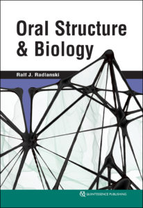
Oral Structure & Biology

Ralf J. Radlanski

Knowledge of the structures of the orofacial region from the macroscopic scale to the molecular level and pathologic changes to those structures enables practitioners to successfully treat patients or seek treatment options. This book presents the structural biologic foundations underpinning dental and oral medicine. Beginning with an overview of the anatomy of the mouth and moving on to the evolution of the oral structures and pre- and postnatal development of the oral cavity, related facial structures, and the teeth, this book describes each part of the orofacial region in terms of its morphology, tissue structure, cellular properties, and development. Functioning as both a textbook for dental students and a reference manual for experienced clinicians, this compendium of the structural biologic foundations of clinical work in dental and oral medicine allows practitioners to integrate current research in molecular biology into a solid framework of knowledge.

472 pp (softcover); 245 illus; ©2018; ISBN 978-0-86715-746-8 (B7468); Now available! $168


Clear Aligner Technique

Sandra Tai

Clear aligners are the future of orthodontics, but digital orthodontics evolves so rapidly that it is hard to keep pace. This book approaches clear aligner treatment from a diagnosis and treatment-planning perspective, discussing time-tested orthodontic principles like biomechanics and anchorage and demonstrating how to apply them to orthodontic cases using these appliances. Each chapter explains how to use clear aligners to treat a given malocclusion and teaches clinicians how to program a suitable treatment plan using available software, how to design the digital tooth movements to match the treatment goals, and finally how execute the treatment clinically and finish the case well. This clinical handbook will prepare orthodontists and dental students to exceed patient expectations with the most esthetic orthodontic appliance currently available.

320 pp; 1,344 illus; ©2018; ISBN 978-0-86715-777-2 (B7772); Now available! $218

Read more about Clear Aligner Technique here!

Clear Aligner Technique: Bringing Orthodontic Treatment into the Digital Era


Current Issues in Journals


prd_banner

Featured article: The Laterally Closed Tunnel for the Treatment of Deep Isolated Mandibular Recessions: Surgical Technique and a Report of 24 Cases
Anton Sculean and Edward P. Allen

A 13- to 32-Year Retrospective Study of Bone Stability for Machined Dental Implants
Massimo Simion, Myron Nevins, Giulio Rasperini, and Francesco Tironi

Immediate Implant Placement in Single-Tooth Molar Extraction Sockets: A 1- to 6-Year Retrospective Clinical Study
Francesco Amato and Giorgio Polara

ijp_banner

Prevalence of Abnormal Morphology of the Temporomandibular Joint in Asymptomatic Subjects: A Retrospective Cohort Study Utilizing Cone Beam Computed Tomography
Thiago Azário de Holanda, Rita de Cássia Costa Ribeiro de Almeida, Alexandre Emídio Ribeiro Silva, Melissa Feres Damian, and Noéli Boscato

Peri-implant Tissue Health and Bone Resorption in Patients with Immediately Loaded, Implant-Supported, Full-Arch Prostheses
Maria Menini, Paolo Setti, Paolo Pera, Francesco Pera, and Paolo Pesce

Investigation of Clinical and Laboratory Wear in Locator-Supported, Implant-Retained Overdentures
Sebastian Hahnel, Christos Alamanos, Sibylle Schneider-Feyrer, Matthias Stöckle, and Martin Rosentritt

jomi_banner

Featured Article: Impact of Surface Chemistry Modifications on Speed and Strength of Osseointegration
Hyeon-Goo Kang, Yong-Soo Jeong, Yoon-Hyuk Huh, Chan-Jin Park, and Lee-Ra Cho

Thematic Abstract Review: Dental Implant Treatment of Atrophic Arches With or Without Augmentation
Jan-Eirik Ellingsen

Evaluation of Two 3D Printers for Guided Implant Surgery
Bruno Sommacal, Miodrag Savic, Andreas Filippi, Sebastian Kühl, and Florian M. Thieringer

banner_ofph

Editorial: A Parting Perspective of the Journal’s Evolution
Barry J. Sessle

Peripheral Glial Cell Line–Derived Neurotrophic Factor Facilitates the Functional Recovery of Mechanical Nociception Following Inferior Alveolar Nerve Transection in Rats
Masahiro Watanabe, Masamichi Shinoda, Dulguun Batbold, Naoyuki Sugano, Shuichi Sato, and Koichi Iwata

Analgesic Effects of Intranasal Ketamine in Rat Models of Facial Pain
Rafaela Claudino, Carina Nones, Erika Araya, and Juliana Chichorro

Short clinical screening procedure for initial diagnosis of temporomandibular disorders
Georg Meyer

Multidisciplinary treatment-increase of vertical dimension combined with Invisalign treatment
Beatriz Solano Mendoza, Lorena Gómez García, Hourieh Pourhamid, and Enrique Solano

Correcting severe deep bite with the Invisalign appliance
Bärbl Reistenhofer, Fanny Triessnig, and Katharina Besser


Dental Meetings Quintessence Will Attend in August


Dental XP Implant Summit
hosted by Dental XP, August 11–12 in New York, New York

XXIII Seminario Ivoclar Vivadent: #E21
hosted by Ivoclar Vivadent, August 17–18 in Ciudad de México, México


Upcoming Quintessence Events


Posted in Books, Journals, Multimedia, Promotions, Roundup, Special Offer, What's New | Leave a comment

Quintessence Roundup: July

Reading time: 9 minutes

July Monthly Special


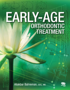
Early-Age Orthodontic Treatment

Aliakbar Bahreman

This book presents readers with the information necessary to understand the morphogenesis of orthodontic problems, to differentiate among various conditions, and to apply early intervention approaches to optimal effect. Through integration of basic science and clinical practice, detailed case reports, and abundant illustrations, the author provides an overview of dental development, from tooth formation to permanent occlusion; emphasizes the developmental stages that must be recognized during patient examination to facilitate differential diagnosis; presents clear, step-by-step instructions for different treatment options; and demonstrates the benefits achieved by intervention in developing malocclusions and guidance of eruption. The discussions in part 1 of the concept of early-age treatment and the basic foundation of occlusal development empower practitioners to detect anomalies and intervene as necessary. Part 2 consists of chapters explaining the ontogeny, diagnosis, and early detection of, as well as the proper intervention for, nonskeletal problems. Part 3 includes chapters on early intervention for dentoskeletal problems in the sagittal, transverse, and vertical dimensions. This book cuts through the controversy surrounding early versus late treatment and shows that clinicians must decide on a case-by-case basis when to provide orthodontic treatment.

436 pp; 1,534 illus; ©2013; ISBN 978-0-86715-566-2 (B5662); $142 Special price! $78


New Titles in Books


Cephalometry in Orthodontics: 2D and 3D

Edited by Katherine Kula and Ahmed Ghoneima

Cephalometrics has been used for decades to diagnose orthodontic problems and evaluate treatment. However, the shift from 2D to 3D radiography has left some orthodontists unsure about how to use this method effectively. This book defines and depicts all cephalometric landmarks on a skull or spine in both 2D and 3D and then identifies them on radiographs. Each major cephalometric analysis is described in detail, and the linear or angular measures are shown pictorially for better understanding. Because many orthodontists pick specific measures from various cephalometric analyses to formulate their own analysis, these measures are organized relative to the skeletal or dental structure and then compared or contrasted relative to diagnosis, growth, and treatment. Cephalometric norms (eg, age, sex, ethnicity) are also discussed relative to treatment and esthetics. The final chapter shows the application of these measures to clinical cases to teach clinicians and students how to use them effectively. As radiology transitions from 2D to 3D, it is important to evaluate the efficacy and cost-effectiveness of each in diagnosis and treatment, and this book outlines all of the relevant concerns for daily practice.

208 pp; 338 illus;©2018; ISBN 978-0-86715-762-8 (B7628); $118 Special preorder price! $94
Available Summer 2018


Short and Ultra-Short Implants

Edited by Douglas Deporter

Research has shown that short implants are not only a viable option but oftentimes a superior one that carries fewer risks for the patient and dentist, especially in resorbed jaw sites. As clinical trials continue to underscore the safety and efficacy of short implants, more dentists are considering their use with real interest, and this book provides the information clinicians need to incorporate short implants into their own practice. The book reviews the clinical effectiveness of short implants and then describes treatment protocols for the various types of short implants and their placement in different areas of the mouth. Case presentations demonstrate the recommended techniques and showcase the results.

168 pp; 334 illus; ©2018; ISBN 978-0-86715-785-7 (B7857); $85 Special preorder price! $68
Available Summer 2018


Botulinum Toxin for Facial Harmony

Altamiro Flávio

The mastery of dentistry brings esthetic knowledge of the face that is applicable to more than just the teeth. In the process of performing a complete facial analysis, the practitioner can identify asymmetries and concerns localized to an area—such as the forehead, eyebrows, nose, or lower face—and offer Botox therapy to increase facial harmony. This book outlines the many clinical uses for Botox, with detailed illustrations and case presentations to support each procedure. The first part of the book covers systematic facial analysis, photographic documentation, and how to plan treatment. Special attention is paid to the anatomy and physiology of the face and the identification of injection points. Detailed treatment instructions for dosage, syringe type, and needle size are included for each procedure, as well as guidelines on how to evaluate results anthropometrically to determine whether esthetic treatment goals have been met. This stunning book will change the way you approach facial analysis and widen your esthetic treatment options for patients.

160 pp; 359 illus; ©2018; ISBN 978-0-86715-787-1 (B7871); Available now! $148

Read more about Botulinum Toxin for Facial Harmony here!

Botulinum Toxin for Facial Harmony: How this Unconventional Treatment Can Expand Your Practice


Oral Structure & Biology

Ralf J. Radlanski

Knowledge of the structures of the orofacial region from the macroscopic scale to the molecular level and pathologic changes to those structures enables practitioners to successfully treat patients or seek treatment options. This book presents the structural biologic foundations underpinning dental and oral medicine. Beginning with an overview of the anatomy of the mouth and moving on to the evolution of the oral structures and pre- and postnatal development of the oral cavity, related facial structures, and the teeth, this book describes each part of the orofacial region in terms of its morphology, tissue structure, cellular properties, and development. Functioning as both a textbook for dental students and a reference manual for experienced clinicians, this compendium of the structural biologic foundations of clinical work in dental and oral medicine allows practitioners to integrate current research in molecular biology into a solid framework of knowledge.

472 pp (softcover); 245 illus; ©2018; ISBN 978-0-86715-746-8 (B7468); Now available! $168


Clear Aligner Technique

Sandra Tai

Clear aligners are the future of orthodontics, but digital orthodontics evolves so rapidly that it is hard to keep pace. This book approaches clear aligner treatment from a diagnosis and treatment-planning perspective, discussing time-tested orthodontic principles like biomechanics and anchorage and demonstrating how to apply them to orthodontic cases using these appliances. Each chapter explains how to use clear aligners to treat a given malocclusion and teaches clinicians how to program a suitable treatment plan using available software, how to design the digital tooth movements to match the treatment goals, and finally how execute the treatment clinically and finish the case well. This clinical handbook will prepare orthodontists and dental students to exceed patient expectations with the most esthetic orthodontic appliance currently available.

320 pp; 1,344 illus; ©2018; ISBN 978-0-86715-777-2 (B7772); Now available! $218

Read more about Clear Aligner Technique here!

Clear Aligner Technique: Bringing Orthodontic Treatment into the Digital Era


Orofacial Pain: Guidelines for Assessment, Diagnosis, and Management

American Academy of Orofacial Pain
Edited by Reny de Leeuw and Gary D. Klasser

Following in the tradition of the previous editions, this book offers the latest research and most up-to-date information on orofacial pain, including a concise overview of each condition as well as its symptoms, comorbidities, differential diagnosis, and treatment options. Every chapter has undergone critical updates to reflect the developments in the expanding field of orofacial pain, including the glossary. These updates include the addition of new diseases such as first-bite syndrome, revised information on genetic factors to reflect new insights gleaned from the OPPERA studies, expanded information on management strategies for certain conditions, and revisions to screening tools for biobehavioral factors. In addition, the classification system used in the fifth edition has been replaced and extended and includes DSM-5 codes where applicable. This book remains an essential tool for any health care provider who treats patients with orofacial pain.

336 pp (softcover); 9 illus; ©2018; ISBN 978-0-86715-768-0 (B7680); Now available! $56

Read more about Orofacial Pain: Guidelines for Assessment, Diagnosis, and Management here!

Orofacial Pain Management in Dentistry: Three Decades of the AAOP Guidelines


Dentistry and the Pregnant Patient

Daniel Ninan

To err on the side of caution, some dentists may hesitate to perform necessary procedures on pregnant patients. However, good oral health is essential to a healthy pregnancy and can help reduce prenatal complications, including preterm delivery and fetal loss. This book serves as an easy-to-use guide to help dentists of all specialties provide safe, effective care for their patients during pregnancy. The book is broken down into chapters on general treatment guidelines, pregnancy-related conditions in each organ system that may impact care, and the safety of the use of common dental drugs, including anesthetics, during pregnancy. With over 50 quick-reference charts and tables and a breakdown of treatment recommendations by trimester, this book is a must-have for any dental office.

160 pp (softcover); ©2018; ISBN 978-0-86715-779-6 (B7796); Now available! $48

Read more about Dentistry and the Pregnant Patient here!

Dental Treatment for Pregnant Patients: What’s Holding Us Back?


Current Issues in Journals


prd_banner

Featured article: The Laterally Closed Tunnel for the Treatment of Deep Isolated Mandibular Recessions: Surgical Technique and a Report of 24 Cases
Anton Sculean and Edward P. Allen

A 13- to 32-Year Retrospective Study of Bone Stability for Machined Dental Implants
Massimo Simion, Myron Nevins, Giulio Rasperini, and Francesco Tironi

Immediate Implant Placement in Single-Tooth Molar Extraction Sockets: A 1- to 6-Year Retrospective Clinical Study
Francesco Amato and Giorgio Polara

ijp_banner

Prevalence of Abnormal Morphology of the Temporomandibular Joint in Asymptomatic Subjects: A Retrospective Cohort Study Utilizing Cone Beam Computed Tomography
Thiago Azário de Holanda, Rita de Cássia Costa Ribeiro de Almeida, Alexandre Emídio Ribeiro Silva, Melissa Feres Damian, and Noéli Boscato

Peri-implant Tissue Health and Bone Resorption in Patients with Immediately Loaded, Implant-Supported, Full-Arch Prostheses
Maria Menini, Paolo Setti, Paolo Pera, Francesco Pera, and Paolo Pesce

Investigation of Clinical and Laboratory Wear in Locator-Supported, Implant-Retained Overdentures
Sebastian Hahnel, Christos Alamanos, Sibylle Schneider-Feyrer, Matthias Stöckle, and Martin Rosentritt

jomi_banner

Featured Article: Influence of Alveolar Bone Defects on the Stress Distribution in Quad Zygomatic Implant–Supported Maxillary Prosthesis
Yuanyuan Duan, Ravi Chandran, and Denise Cherry

Thematic Abstract Review: The Implant-Abutment–Soft Tissue Interface: Change Is Hard!
Thomas W. Oates

Clinical Outcomes of Vertical Distraction Osteogenesis for Dental Implantation: A Systematic Review and Meta-Analysis
Kai Zhao, Feng Wang, Wei Huang, and Yiqun Wu

banner_ofph

Shared Genetics of Temporomandibular Disorder Pain and Neck Pain: Results of a Twin Study
Corine M. Visscher, Maarten J. Schouten, Lannie Ligthart, Caroline M.H.H. van Houtem, Ad de Jongh, and Dorret I. Boomsma

Predictors of Long-Term Temporomandibular Disorder Pain Intensity: An 8-Year Cohort Study
Flavia P. Kapos, John O. Look, Lei Zhang, James S. Hodges, and Eric L. Schiffman

Effects of a Physical Therapy Protocol in Patients with Chronic Migraine and Temporomandibular Disorders: A Randomized, Single-Blinded, Clinical Trial
Miriam Garrigós-Pedrón, Roy La Touche, Pablo Navarro-Desentre, Manuel Gracia-Naya, and Eva Segura-Ortí

Evaluation of Invisalign treatment using the Peer Assessment Rating (PAR) index
Maria del Mar Ramis, Vicente Gandia, Carlos Bellot-Arcís, Rosa Cibrian, Vanessa Peredes-Gallardo, and Jose Luis Gandia

Assessment of phonetic alterations in patients treated with F22 aligners
Angela Arreghini, Arianna Beretta, Luca Lombardo, Claudia Almoni, and Giuseppe Siciliani

Effect of composite attachment on initial force system generated during canine rotation with plastic aligners: A three-dimensional finite elements analysis
Juan Pablo Gómez Arango, Fabio Marcelo Peña, Esteban Valencia, and Carlos Eduardo Mesa


Upcoming Quintessence Events


Posted in Books, Journal of Oral & Facial Pain and Headache, Journals, Promotions, Roundup, Special Offer, The International Journal of Oral & Maxillofacial Implants, The International Journal of Periodontics & Restorative Dentistry, The International Journal of Prosthodontics, What's New | Tagged , , | Leave a comment

Botulinum Toxin for Facial Harmony: How this Unconventional Treatment Can Expand Your Practice

Reading time: 12 minutes

Dentists are known for their pursuit of perfection. It’s one of the traits that define the profession; arguably, the only trait more significant is their desire to help and serve their patients. Altamiro Flávio, DDS, author of a new clinical book Botulinum Toxin for Facial Harmony, is a dentist who possesses both of these traits. Twelve years ago, he discovered a treatment that could greatly benefit his patients, but he lacked the education and experience to provide it. In order to meet this patient need, he embarked on an educational journey that would drastically change his practice philosophy and the course of his career.

The Importance of the Face in Dentistry

So many of the treatment-planning decisions that dentists make involve careful consideration of the face. A good clinician uses reference points of the face when planning restorative or prosthodontic treatments; further, an esthetic result relies on the clinician’s ability to successfully harmonize the character of the teeth with the character of the face.

The integration of face and smile knowledge will transform the way we impact people’s lives.

Christian Coachman, DDS, CDT
“In order to be able to produce esthetic smiles, we must first understand the face itself,” says Christian Coachman, DDS, CDT, founder of Digital Smile Design. “All professionals who work with smile rehabilitation should also be experts on the face. We have noticed a lack of facial knowledge by dentists as well as a lack of knowledge of smiles by plastic surgeons, and this gap should not exist. The integration of face and smile knowledge will transform the way we impact people’s lives by modifying their facial expressions.”

“Early in our college days we learn that we must analyze not just the patient’s mouth but also the face,” Dr Flávio emphasizes. “Thirty years ago, this was easy to say but not feasible to do. These days we have facial references and concepts like Digital Smile Design that use facial aspects to build, reshape, and position the teeth. But imagine if the facial aspects that we use as reference points are not adequate. What can we as dentists do?”

The answer to this question isn’t as complicated as you may think, and it comes in a little glass vial.

“In 2006, I participated in a smile competition sponsored by the Brazilian Society of Aesthetic Dentistry,” Dr Flávio recalls. “During this kind of competition, some very important dentists judge before-and-after photographs from cases of dental work related to esthetics. During the prequalification process, one of the judges told me that although my work was beautiful, the lips of the patient showed some asymmetry, so I probably wouldn’t win. I ended up winning third place, and I then referred the patient in this case to a plastic surgeon, Dr Marcio Rocha, for surgery to fix the asymmetry.”

But during the consult, Dr Marcio told him that surgery was not an option: the best way to fix the asymmetry was botulinum toxin treatment. By observing the treatment and its results, Dr Flávio realized this was a fast, precise, minimally invasive, and reversible procedure, and he wanted to be able to offer it to his patients. So he took his first course at the renowned Ligia Kogos Dermatology Clinic in São Paulo, and thus began his journey with botulinum toxin treatment in dentistry.

(a and b) Before and after treatment for asymmetric smile. This patient’s buccal corridor and commissure asymmetries were treated using botulinum toxin.

Who Treats the Face

Countless other dental clinicians have also found ways to implement botulinum toxin treatment into their practices in ways that enhance and augment dental treatments. But is this overstepping? Is the face part of dentistry’s jurisdiction, or should it be left to a plastic surgeon or dermatologist? Why should dentists treat facial concerns?

The mouth is part of the face; it should not be evaluated separately.

Altamiro Flávio, DDS
“There are some people who say that dentists should only be allowed to treat the perioral area while dermatologists should treat the rest of the face,” Dr Flávio says. “I disagree because of the risk created when botulinum toxin treatment is managed by multiple clinicians. Because of immunogenicity, a patient must wait 4 months between botulinum toxin treatments. If a patient receives another round of treatment before that window has passed—perhaps because they are being treated by a dermatologist for wrinkles while also receiving anti-bruxism treatment from a dentist—it will provoke a vaccine effect from the patient’s immune system and decrease the action of the botulinum toxin in subsequent treatments.”

Treating areas so close to each other with different professionals may also interfere with the final result of both treatment plans because of how areas of the face affect each other. Dr Flávio stresses that the decision to be treated by a dentist or by a physician is up to the patient—the important thing is that it be one or the other, not both. Nonetheless, while a dentist possesses the foundational knowledge to learn botulinum toxin treatments for the face, dermatologists lack the dental knowledge required to provide structural and therapeutic treatments for the mouth. Dentists are also uniquely equipped to manage esthetic harmony and integration between the mouth and the face.

“It is not possible to delegate the mouth to physicians,” Dr Flávio stresses, “because they are more versed in anti-aging treatment for the mouth, and their main treatment focus is often the upper third of the face. With the treatment options we have today, including botulinum toxin, we as dentists can address those esthetic and functional facial issues. The mouth is part of the face; it should not be evaluated separately.”

Teaching the Science Behind Facial Esthetics

In order to spread awareness of the possible applications of botulinum toxin in dentistry, Dr Flávio developed a continuing education course called “A Smile for Each Face,” which combines dental topics such as facial analysis, digital planning, dental anatomy, direct and indirect veneers, and restorations with dermatologic topics like botulinum toxin, facial fillers, and viscosupplementation. He has since taught on these topics in 6 different countries, helping others to expand the treatment options they can offer their patients.

“During my courses,” Dr Flávio explains, “students learn the name, function, and position of each facial muscle. As a teacher, I know that visualization is a very strong component of the learning process. Some time ago, I was at a birthday party where I saw children running around with butterflies and other animals painted on their faces. I realized I could use this technique as a learning aid for my students. So at the end of my courses, in order to complete the learning process my students must be able to draw the muscles on someone’s face. It’s a way to own the knowledge. It wouldn’t work if the drawing was made over a photograph of a face, as the muscles should be located by considering the palpation of muscles and bones and also the muscular activity. Similarly, injections are not made on a photograph—they are made directly on the patient’s face.”

Dr Flávio’s painting exercise brings to the surface the muscular structures that the students will need to locate and treat in their patients, forming a direct connection between theoretical knowledge and practice. Location is paramount for successful botulinum toxin treatment, and the painting teaches students how to visualize the current state of the patient’s muscles and imagine the results of potential treatments. They are then able to reimagine the patient’s face and work to achieve those treatment goals. But what exactly can botulinum toxin treatment accomplish in dentistry?

Applications of Botulinum Toxin in Dentistry

The main benefit of botulinum toxin in a dental practice is to provide a more comprehensive treatment plan for your patients. So it makes sense to consider both therapeutic and structural benefits alongside esthetic ones.

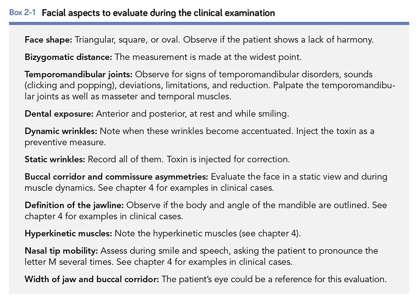
In his book Botulinum Toxin for Facial Harmony, Dr Flávio breaks down the steps of this treatment-planning process in a concept called facial analysis. By using mathematic rules to analyze the face, clinicians can identify facial issues as well as reasons to improve those esthetic issues.

Esthetics is an objective topic, not a subjective one.

Altamiro Flávio, DDS
“When we use the rules of facial analysis to evaluate our patients’ faces scientifically, we realize that esthetics is indeed an objective topic rather than a subjective one.” Dr Flávio explains. “During the first appointment, dentists should analyze the patient’s face and identify all possible issues, not only around the mouth but across the whole face,” he says. “When presenting the diagnosis and treatment plan to the patient, the dentist should never split the procedures into ‘dental’ and ‘facial.’ For instance: A patient who has dental attrition from bruxism can be treated with Botox injections to decrease the muscle power before the tooth reconstruction is performed. This allows the dentist to first treat the cause (bruxism) and then the consequence (dental attrition). If this same patient has wrinkles and an inadequately positioned eyebrow, the treatment plan should address those issues as well so that the patient receives therapeutic, anti-aging, and structural benefits all from the same treatment plan. Many books on this topic tell you how to do these procedures, but none of them tell you why to do it. The reader will find the reasons for esthetic treatments in my book.”

(a to f) This patient’s dynamic wrinkles around the eyes were beginning to set in as static wrinkles, so they were treated with botulinum toxin. Her gummy smile was also treated with an injection in the depressor septi nasi muscle, further reducing the wrinkles at the eyes. The final view shows markedly fewer dynamic wrinkles and resolution of the gummy smile (f).

(a to e) This patient’s periorbital wrinkles were present even when the face was at rest, so treatment with botulinum toxin was performed to smooth them. (f and g) Restorations were placed on the maxillary anterior teeth to elongate the crowns and give the face a more dolichofacial appearance. (h and i) Comparison of pretreatment and posttreatment smiles. Note the younger-looking appearance after treatment.

(a and c) Pretreatment views. In the upper lip, the levator labii superioris alaeque nasi and levator labii superioris were hyperkinetic, excessively elevating the upper lip. Besides the gummy smile, another point of concern for the patient was the intense closing of her eyes when she smiled. (b and d) Final views 10 days after BTX injection. (e) Injection points. Botulinum toxin was used to promote the chemodenervation of the muscles that elevate the upper lip and depress the lower lip during the smile. This reduced the movement of the lips during the smile and resulted in less exposure of the maxillary gingival tissue. In this specific patient, the gummy smile could not be treated through periodontal plastic surgery (crown lengthening) because the clinical crown ratio was already ideal, harmonious, and esthetically pleasant.

(a) Pretreatment view of a patient with a visually wide lower third. The patient complained of preauricular pain when waking up, located at the masseter (left and right), with no tension headache. She also reported diurnal and nocturnal teeth clenching. Signs and symptoms included enamel cracks, dental attrition, limited mouth opening (35 mm), clicking in the right temporomandibular joint with reduction, and maxillary and mandibular bone exostoses. The patient was diagnosed with exaggerated, abnormal, and continued tension or contraction of the mandibular elevator muscles peculiar to bruxism. (b) The facial analysis identified apparent hypertrophy in the masseter muscles because the bigonial distance was almost the same as the bizygomatic distance. The top dotted line measures the bizygomatic distance, and the bottom dotted line shows the bigonial distance. The triangle base shows the difference in width between the two measurements. The shorter this distance, the greater the masseter hypertrophy. Botulinum toxin was used to promote the chemodenervation of the masseter muscles (left and right) and the temporalis muscles (left and right), adjusting their excessive contraction. (c and d) Comparison of the pretreatment condition and the result 30 days after the first toxin injection. The triangles in both photographs show the amount of narrowing in the lower third of the face after BTX injection. (e and f) Anthropometrics showing a 14-mm narrowing of the lower third 30 days after the injection.

The procedures described in Dr Flávio’s book are organized by individual muscle, and all information required for the treatment—anatomical details, instructions for locating the muscle, injection protocol, and safety warnings—is provided in the same place to increase the efficacy of the book as a clinical resource. Illustrations show the location of the muscle both in a cadaver and in a patient, and videos that can be accessed via QR code show how to locate the injection points for each muscle. Detailed treatment instructions for dosage, syringe type, and needle size are included for each procedure, as well as guidelines on how to evaluate results anthropometrically to determine whether esthetic treatment goals have been met.

Pages from Dr Flávio’s book. The procedures in his book are organized by individual muscle, with illustrations showing the location of the muscle in a patient and in a cadaver. Detailed instructions for dosage, syringe type, and needle size are also provided.

In cases where dentistry has already established traditional solutions, such as a gummy smile, dentists may hesitate before considering a treatment that necessitates repetition. There are advantages and disadvantages to every treatment. In many situations, the advantages of botulinum toxin as a minimally invasive, reversible treatment may outweigh the perceived disadvantage of its duration of effectiveness.

“My students often complain that botulinum toxin treatments only last 5 months,” Dr Flávio says. “When they do this, I ask them to name one antibiotic, anti-inflammatory, anesthetic, or antidepressant that has an effect that lasts as long. When we think of it this way, the duration time becomes one of the greatest advantages of the toxin.”

The use of botulinum toxin expands the range of what dentists can treat for their patients. With this tool, dentists can better serve their patients with esthetic facial concerns not only by correcting what’s inside the mouth, but also by adjusting the overall presentation of the face.

“I get excited when I see the final results of treatment in my daily practice,” Dr Flávio emphasizes. “This professional accomplishment is a great reward. These days, the world experiences very fast development in medical areas. All professionals must evolve to absorb these changes, and dentists are no different. With botulinum toxin, we now have a great opportunity to use the knowledge we learned when studying anatomy and physiology in dental school by spreading the benefits of botulinum toxin treatment to the perioral region and the facial area. I incorporated the use of botulinum toxin into my daily practice not because it’s a hot topic, but to address my patients’ needs. Now, I can’t see myself working without botulinum toxin in my office.”


Altamiro Flávio, DDS, graduated from the Federal University of Goiás College of Dentistry in 1990 and went on to become a specialist in dental prostheses at the Federal University of Uberlândia in 1992. In 2010, he created a continuing education course called “A Smile for Each Face,” which included topics such as facial analysis, digital planning, dental anatomy, direct and indirect veneers, restorations, botulinum toxin, facial fillers, and viscosupplementation. He continues to teach courses like this all over the world, and he also teaches the specialization course in restorative dentistry at the Brazilian Association of Dentistry in Goiás. Dr Flávio is an accredited member of the Brazilian Society of Aesthetic Dentistry and a founder of the Brazilian Society of Botulinum Toxin and Facial Implants. He maintains a private practice in Goiânia, Brazil.

 

Botulinum Toxin for Facial Harmony

Altamiro Flávio

The mastery of dentistry brings esthetic knowledge of the face that is applicable to more than just the teeth. In the process of performing a complete facial analysis, the practitioner can identify asymmetries and concerns localized to an area—such as the forehead, eyebrows, nose, or lower face—and offer Botox therapy to increase facial harmony. This book outlines the many clinical uses for Botox, with detailed illustrations and case presentations to support each procedure. The first part of the book covers systematic facial analysis, photographic documentation, and how to plan treatment. Special attention is paid to the anatomy and physiology of the face and the identification of injection points. Detailed treatment instructions for dosage, syringe type, and needle size are included for each procedure, as well as guidelines on how to evaluate results anthropometrically to determine whether esthetic treatment goals have been met. This stunning book will change the way you approach facial analysis and widen your esthetic treatment options for patients.

160 pp; 359 illus; ©2018; ISBN 978-0-86715-787-1 (B7871); Available now! $148

Posted in Books, Esthetic Dentistry, Feature, Misc, Multidisciplinary, What's New | Tagged , , , , , , | 5 Comments

Quintessence Roundup: June

Reading time: 9 minutes

June Monthly Special


Defense From Within: A Guide to Success As a Dental Malpractice Defense Expert

Jeffrey A. Krompier

In this straightforward and highly informative book, an attorney with nearly 30 years of experience defending health care providers against malpractice suits offers dental clinicians essential advice and inside information regarding what it takes to be a successful defense expert witness. Those interested in taking on this challenge will discover the characteristics, skills, and assets that are required and determine whether they are well suited for the role. From there, the text details the work involved in serving as an expert witness, such as case assessments and literature reviews, expert opinions, deposition testimony, and handling examination in the courtroom. Finally, the author offers helpful tips on building a curriculum vitae and marketing oneself as a defense expert witness, thereby providing the reader with all the tools required to enter into this unique service well prepared for success.

144 pp (softcover); ©2012; ISBN 978-0-86715-583-9 (B5839); $42 Special price! $2


New Titles in Books


Botulinum Toxin for Facial Harmony

Altamiro Flávio

The mastery of dentistry brings esthetic knowledge of the face that is applicable to more than just the teeth. In the process of performing a complete facial analysis, the practitioner can identify asymmetries and concerns localized to an area—such as the forehead, eyebrows, nose, or lower face—and offer Botox therapy to increase facial harmony. This book outlines the many clinical uses for Botox, with detailed illustrations and case presentations to support each procedure. The first part of the book covers systematic facial analysis, photographic documentation, and how to plan treatment. Special attention is paid to the anatomy and physiology of the face and the identification of injection points. Detailed treatment instructions for dosage, syringe type, and needle size are included for each procedure, as well as guidelines on how to evaluate results anthropometrically to determine whether esthetic treatment goals have been met. This stunning book will change the way you approach facial analysis and widen your esthetic treatment options for patients.

160 pp; 359 illus; ©2018; ISBN 978-0-86715-787-1 (B7871); $148 Special preorder price! $118
Available July 2018

Read more about Botulinum Toxin for Facial Harmony here!

Botulinum Toxin for Facial Harmony: How this Unconventional Treatment Can Expand Your Practice


Oral Structure & Biology

Ralf J. Radlanski

Knowledge of the structures of the orofacial region from the macroscopic scale to the molecular level and pathologic changes to those structures enables practitioners to successfully treat patients or seek treatment options. This book presents the structural biologic foundations underpinning dental and oral medicine. Beginning with an overview of the anatomy of the mouth and moving on to the evolution of the oral structures and pre- and postnatal development of the oral cavity, related facial structures, and the teeth, this book describes each part of the orofacial region in terms of its morphology, tissue structure, cellular properties, and development. Functioning as both a textbook for dental students and a reference manual for experienced clinicians, this compendium of the structural biologic foundations of clinical work in dental and oral medicine allows practitioners to integrate current research in molecular biology into a solid framework of knowledge.

472 pp (softcover); 245 illus; ©2018; ISBN 978-0-86715-746-8 (B7468); Now available! $168


Clear Aligner Technique

Sandra Tai

Clear aligners are the future of orthodontics, but digital orthodontics evolves so rapidly that it is hard to keep pace. This book approaches clear aligner treatment from a diagnosis and treatment-planning perspective, discussing time-tested orthodontic principles like biomechanics and anchorage and demonstrating how to apply them to orthodontic cases using these appliances. Each chapter explains how to use clear aligners to treat a given malocclusion and teaches clinicians how to program a suitable treatment plan using available software, how to design the digital tooth movements to match the treatment goals, and finally how execute the treatment clinically and finish the case well. This clinical handbook will prepare orthodontists and dental students to exceed patient expectations with the most esthetic orthodontic appliance currently available.

320 pp; 1,344 illus; ©2018; ISBN 978-0-86715-777-2 (B7772); Now available! $218

Read more about Clear Aligner Technique here!

Clear Aligner Technique: Bringing Orthodontic Treatment into the Digital Era


Orofacial Pain: Guidelines for Assessment, Diagnosis, and Management

American Academy of Orofacial Pain
Edited by Reny de Leeuw and Gary D. Klasser

Following in the tradition of the previous editions, this book offers the latest research and most up-to-date information on orofacial pain, including a concise overview of each condition as well as its symptoms, comorbidities, differential diagnosis, and treatment options. Every chapter has undergone critical updates to reflect the developments in the expanding field of orofacial pain, including the glossary. These updates include the addition of new diseases such as first-bite syndrome, revised information on genetic factors to reflect new insights gleaned from the OPPERA studies, expanded information on management strategies for certain conditions, and revisions to screening tools for biobehavioral factors. In addition, the classification system used in the fifth edition has been replaced and extended and includes DSM-5 codes where applicable. This book remains an essential tool for any health care provider who treats patients with orofacial pain.

336 pp (softcover); 9 illus; ©2018; ISBN 978-0-86715-768-0 (B7680); Now available! $56

Read more about Orofacial Pain: Guidelines for Assessment, Diagnosis, and Management here!

Orofacial Pain Management in Dentistry: Three Decades of the AAOP Guidelines


Dentistry and the Pregnant Patient

Daniel Ninan

To err on the side of caution, some dentists may hesitate to perform necessary procedures on pregnant patients. However, good oral health is essential to a healthy pregnancy and can help reduce prenatal complications, including preterm delivery and fetal loss. This book serves as an easy-to-use guide to help dentists of all specialties provide safe, effective care for their patients during pregnancy. The book is broken down into chapters on general treatment guidelines, pregnancy-related conditions in each organ system that may impact care, and the safety of the use of common dental drugs, including anesthetics, during pregnancy. With over 50 quick-reference charts and tables and a breakdown of treatment recommendations by trimester, this book is a must-have for any dental office.

160 pp (softcover); ©2018; ISBN 978-0-86715-779-6 (B7796); Now available! $48

Read more about Dentistry and the Pregnant Patient here!

Dental Treatment for Pregnant Patients: What’s Holding Us Back?


Waxing for Dental Students

Rowida Abdalla

When dental students are first taught how to wax teeth, they need clear instructions with logical steps and plenty of illustrations. With the omission of any distracting information about dies, crowns, bridges, and occlusion, this concise book is squarely directed to first-year dental students and can be used as a primary laboratory manual. The author covers the basics of wax instrumentation and addition, but the bulk of the book details the steps for waxing incisors, canines, premolars, and molars. Each chapter includes an introductory waxing exercise for the tooth type, followed by the key points of morphology for the tooth in question. Photographs detail the waxing steps for both a maxillary and a mandibular full crown on a tooth peg and show how the wax-up should mimic the contralateral tooth in terms of dimension, embrasures, heights of contour, line angles, and point angles. The logical and straightforward protocols in this book will help dental students quickly improve their waxing skills and reach expected goals.

88 pp (spiral bound); 388 illus; ©2018; ISBN 978-0-86715-773-4 (B7734); Now available! $58

Read more about Waxing for Dental Students here!

Waxing Poetic: The First Waxing Manual Written Specifically for First-Year Dental Students


Quintessence of Dental Technology 2018

Edited by Sillas Duarte, Jr

QDT 2018 presents original articles with a beautiful array of images showcasing the year’s newest techniques, materials, tools, and innovations. This year’s focus is decidedly digital—digital treatment planning, digital communication, and digital multidisciplinary workflow for esthetic and functional rehabilitations. Minimally invasive techniques are highlighted throughout, including their use for full-mouth rehabilitation and to restore the morphology of teeth damaged by wear. A special “how-to” article on focus stacking macro photography by Carlos Ayala Paz is the first to be published specific to dentistry.

256 pp; 1,090 illus; ©2018; ISBN 978-0-86715-783-3 (J0629); Now available! $148


Esthetic and Restorative Dentistry: Material Selection and Technique, Third Edition

Douglas A. Terry and Willi Geller

Restorative dentistry has seen dramatic advances in recent years, especially with the use of digital technologies, and this book provides the most up-to-date information on enhanced developments, materials, and techniques that have emerged since the publication of the second edition, offering the reader a completely updated, revised, and newly illustrated overview of modern esthetic and restorative dentistry complete with tutorial videos. New topics include web-based communication with the laboratory, indirect composite chairside CAD/CAM restorations, a comparison of digital and conventional techniques, the resin composite injection technique, as well as updated information on composites and ceramic systems, including esthetic zirconia. New cases illustrate the maintenance of esthetic restorative materials, esthetic contouring, immediate dentin sealing, and novel surgical techniques such as lip repositioning, connective tissue grafting, and ridge preservation with collagen membranes. Dr Terry expanded his team of editorial reviewers to include the best minds in research and clinical practice, and the final product is a testament to his dedication to patient satisfaction and treatment success. The techniques demonstrated in this book will no doubt elevate your practice to the next level.

792 pp; 2,584 illus; ©2018; ISBN 978-0-86715-763-5 (B7635); Now available! $348

Read more about Dr Douglas A. Terry and Esthetic and Restorative Dentistry: Material Selection and Technique, Third Edition here!

Author Spotlight: Douglas A. Terry


Current Issues in Journals


prd_banner

Featured article: Increasing the Vertical Dimension of Occlusion: A Multicenter Retrospective Clinical Comparative Study on 100 Patients with Fixed Tooth-Supported, Mixed, and Implant-Supported Full-Arch Rehabilitations
Giacomo Fabbri, Roberto Sorrentino, Giorgio Cannistraro, Francesco Mintrone, Leonardo Bacherini, Roberto Turrini, Tiziano Bombardelli, Michele Nieri, and Mauro Fradeani

Clinical Outcome After 8 to 10 Years of Immediately Restored Single Implants Placed in Extraction Sockets and Healed Ridges
Stefanie Raes, Jan Cosyn, Anabel Noyelle, Filiep Raes, and Hugo De Bruyn

Outcomes After 25 Years of Periodontal Treatment and Maintenance of a Patient Affected by Generalized Severe Aggressive Periodontitis
Marco Clementini, Fabio Vignoletti, and Massimo de Sanctis

ijp_banner

The Effects of Sleeping With or Without Prostheses on Sleep Quality, Sleep Bruxism, and Signs of Obstructive Sleep Apnea Syndrome: A Pilot Study
Paulo Roberto Castro Mattia, Caio Marcelo Panitz Selaimen, Eduardo Rolim Teixeira, Simone Chaves Fagondes, and Márcio Lima Grossi

A Five-Year Retrospective Assay of Implant Treatments and Complications in Private Practice: Restorative Treatment Profiles of Long-Span, Implant-Supported Fixed and Removable Dental Prostheses
Vahed Parzham, Roy B. Judge, and Denise Bailey

Strategic Use of Microscrews for Enhancing the Accuracy of Computer-Guided Implant Surgery in Fully Edentulous Arches: A Case History Report
Du-Hyeong Lee

jomi_banner

Featured Article: Influence of Alveolar Bone Defects on the Stress Distribution in Quad Zygomatic Implant–Supported Maxillary Prosthesis
Yuanyuan Duan, Ravi Chandran, and Denise Cherry

Thematic Abstract Review: The Implant-Abutment–Soft Tissue Interface: Change Is Hard!
Thomas W. Oates

Clinical Outcomes of Vertical Distraction Osteogenesis for Dental Implantation: A Systematic Review and Meta-Analysis
Kai Zhao, Feng Wang, Wei Huang, and Yiqun Wu

banner_ofph

Shared Genetics of Temporomandibular Disorder Pain and Neck Pain: Results of a Twin Study
Corine M. Visscher, Maarten J. Schouten, Lannie Ligthart, Caroline M.H.H. van Houtem, Ad de Jongh, and Dorret I. Boomsma

Predictors of Long-Term Temporomandibular Disorder Pain Intensity: An 8-Year Cohort Study
Flavia P. Kapos, John O. Look, Lei Zhang, James S. Hodges, and Eric L. Schiffman

Effects of a Physical Therapy Protocol in Patients with Chronic Migraine and Temporomandibular Disorders: A Randomized, Single-Blinded, Clinical Trial
Miriam Garrigós-Pedrón, Roy La Touche, Pablo Navarro-Desentre, Manuel Gracia-Naya, and Eva Segura-Ortí

Evaluation of Invisalign treatment using the Peer Assessment Rating (PAR) index
Maria del Mar Ramis, Vicente Gandia, Carlos Bellot-Arcís, Rosa Cibrian, Vanessa Peredes-Gallardo, and Jose Luis Gandia

Assessment of phonetic alterations in patients treated with F22 aligners
Angela Arreghini, Arianna Beretta, Luca Lombardo, Claudia Almoni, and Giuseppe Siciliani

Effect of composite attachment on initial force system generated during canine rotation with plastic aligners: A three-dimensional finite elements analysis
Juan Pablo Gómez Arango, Fabio Marcelo Peña, Esteban Valencia, and Carlos Eduardo Mesa


Dental Meetings Quintessence Will Attend in June


IDIA Summer Symposium
hosted by the International Dental Implant Association, June 2–3 in Chicago, Illinois


Upcoming Quintessence Events


Posted in Books, Journal of Oral & Facial Pain and Headache, Journals, Promotions, Roundup, Special Offer, The International Journal of Oral & Maxillofacial Implants, The International Journal of Periodontics & Restorative Dentistry, The International Journal of Prosthodontics, What's New | Leave a comment