Why Drawing Matters

Reading time: 4 minutes

Dentistry is part science, part craftsmanship, and part artistry. And only when clinicians have skills in all three areas will they produce excellent results and outcomes for their patients. The science and craftsmanship of dentistry are covered extensively in dental school, but the artistry part doesn’t get as much attention. Dr Hanan Elgendy is trying to change that; she teaches her students how to draw the teeth to increase their artistic skill.

In her new book, she explains that “drawing is a skill that can be learned by nearly every person with normal eyesight and average eye-hand coordination.” If you can thread a needle or catch a baseball, you can learn how to draw. She expounds that “learning to draw is more than learning the skill itself; by learning the steps, you will learn how to see…how to put down on paper what you see in front of your eyes,” thereby enhancing your ability to see the fine details in your wax-ups of teeth.
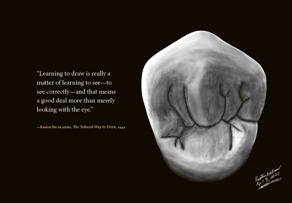
Her method relies on using graph paper and tooth measurements to accurately record the outlines of the five aspects of every tooth—mesial, distal, facial/buccal, lingual, and incisal/occlusal. She writes that “Close observation of the outlines of the squared backgrounds shows the relationship of crown to root, extent of curvatures at various points, inclination of roots, relative widths of occlusal surfaces, height of marginal ridges, contact areas, and so forth.”
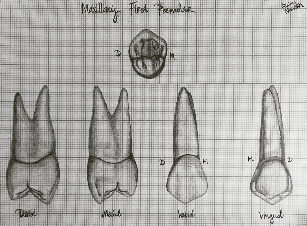
She begins by listing all the measurements of each tooth, including their crown length, root length, mesiodistal and buccolingual diameters of the crown and at the cervix of the crown, and the curvature of the cervical line (mesial and distal). With graph paper, these measurements can be used to draw any tooth in the mouth, and each tooth is given its own unit to show how this is done, followed by a blank page of graph paper to try it out.
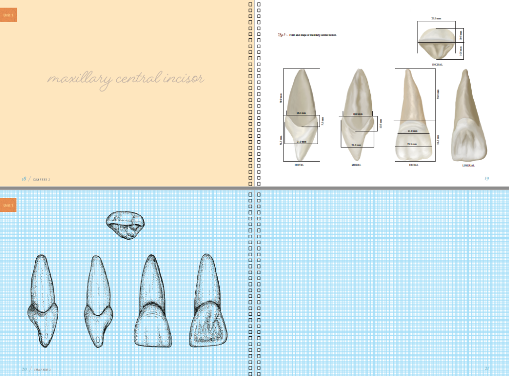
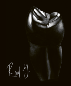
Dr Elgendy knows that drawing is a learnable, teachable skill, because she’s seen the results firsthand. Her students walk into her class with rudimentary skills and leave with the ability to draw this. In fact, nearly all of the book’s illustrations come from her students. And that’s why she is committed to teaching as many dentists as she can how to draw. Because drawing trains the eyes to see the teeth and therefore teaches you how to capture their details first on paper and then in your restorative work. Ask any master like Pascal Magne, and they will tell you that the artistry matters just as much as the materials you use. If you want to improve your artistic eye or if you simply enjoy drawing and want to take that interest to the next level, this book is perfect for you. Preview How to Draw Teeth and Why It Matters here.


Hanan Elgendy, BDS, MS, is Clinical Assistant Professor in the Department of General Dentistry, Operative Dentistry Division at East Carolina University (ECU) School of Dental Medicine in Greenville, North Carolina, where she is the director of the Dental Anatomy, Dental Photography, and Advanced Esthetic Operative courses. Dr Elgendy earned her BDS from Ain Shams University in Egypt and was in private practice for 5 years before moving to the United States in 2012. She completed a master’s degree in operative dentistry at the University of Iowa College of Dentistry in 2016 and was a clinical assistant professor in the Department of Operative Dentistry there until joining ECU in 2019. Her major interest is enhancing student learning in dental anatomy, dental photography, CAD/CAM, and cosmetic dentistry. Dr Elgendy has dedicated her career to teaching restorative dentistry, and she takes pride in knowing how much her students love learning how to draw teeth.
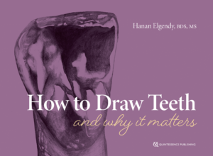
How to Draw Teeth and Why It Matters
Hanan Elgendy

Understanding tooth morphology and anatomical form is crucial to being a good dentist to ensure both function and esthetics. Dr Elgendy contends that the ability to draw an accurate outline of a tooth is a good indication that a student has clearly seen and understood its external morphology. After all, learning to draw the fine details of the tooth is really learning to see them in the first place. That’s why she created this book to guide dental students to seeing and reproducing tooth morphology. The workbook begins with the basics of drawing and quickly shifts to the details of each tooth and how to tackle its morphology. Practice pages are included for each tooth, with extra pages at the end for further practice. Part art book and part workbook, this beauty will inspire students and dentists alike to see better and capture what they see in their restorations.

120 pp (spiral bound); 160 illus; ©2022; ISBN 978-1-64724-0-48 (B0448); US $48

Posted in Multidisciplinary, Restorative Dentistry, What's New | Leave a comment

Complete Denture Therapy Made Simple

Reading time: 7 minutes

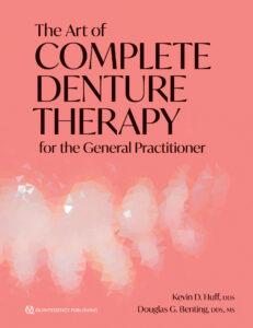
It is no secret in the dental community that dental implants are seen as the best choice for tooth replacement. But not everyone can have dental implants for a variety of reasons. Many people still require traditional dentures, but most practitioners don’t feel confident making them. In their new book, experienced clinicians Drs Kevin Huff and Doug Benting show that complete dentures don’t have to be cumbersome or scary. The Art of Complete Denture Therapy for the General Practitioner is a thorough but concise guide to providing complete denture care from start to finish—without all of the boring stuff from classic textbooks! In fact, they include many pragmatic and cost-saving tips and tricks using unconventional tools to get the job done. Let’s look at some of their most used materials that you may be more likely to find at your local hardware store than from a dental supplier.

Play-Doh and more

You might have heard it before—colorful children’s modeling compound has a surprising number of uses in dentistry. And why not? It’s inexpensive, versatile, eye-catching, safe, and sometimes reusable depending on the application. Dr Huff uses it for blocking out undercuts in the mouth under implants and bridges when making an impression because it is water soluble and nontoxic. In denture fabrication, it can be used for boxing to form the land areas of master gypsum casts because it can be easily removed from the stone with a toothbrush and water.

Play-Doh can also be used to block out undercuts in a denture for remounting and repair  procedures.  As Dr Huff explains, “Before Play-Doh for boxing, I was taught to use a mix of pumice and quick-set stone in a rubber mixing bowl as the matrix for boxing. That took more time to set up and was a mess to work with.” Play-Doh, on the other hand, comes already set and tends to stick to itself while being easily moldable. It can be trimmed or pressed to create a seal between the frame and impression during boxing before the stone is poured, and is easily cleaned off when the master cast is ready to be separated from the base.

Fig 1 Boxed impression with Play-Doh and duct tape.

When using Play-Doh for boxing, it can be trimmed with a cake icing spatula, a table knife, or a lab knife. Then, the eraser end of a pencil or even a fingertip can be used to shape the Play-Doh to the impression. Tried the Play-Doh technique and have some excess when separating the master cast? An extra toothbrush can be wet and used to clean off the cast.

Fig 2 Using a pencil to shape the Play-Doh.

Corrected graduated cylinder

When you want an exact measurement of water or other liquid, a graduated cylinder can be your friend. But when you need this measurement to be consistently precise, you run the risk of over- or underpouring by a little bit or of getting a distorted view of the volume by looking at the cylinder from the wrong angle.  Here’s a pearl for predictability: cut a hole at the measurement needed with a diamond disk. In this way, any excess water will drain out, providing you with exactly the volume the recipe calls for every time.

Fig 3 A corrected graduated cylinder allows excess water to drain out, allowing for more precise measurements.

Duct tape and ceramic tile

Duct tape serves as a great option for boxing a cast. Drs Huff and Benting use either duct tape or magnetic boxing strips during the box and pour technique. The authors explain, “The advantage of using duct tape is cost and convenience, but the tape is not reusable. Magnetic strips are inexpensive and can be reused many times; however, if the perimeter of the impression and Play-Doh base is greater than the length of the magnetic strips, they are not useful, and the duct tape method is more applicable.”

Along with the duct tape, a small ceramic wall tile, eg, 4 × 4 inches, can be used as a surface to perform the boxing technique. It is a good thickness for handling and is portable, reusable, and inexpensive.

Fig 4 Impression and cast on a tile with magnetic strips.

Making wax rims for the record base

When you need a warm surface to shape the wax, make your own hot plate! Have a paint scraper or metal putty knife? A Bunsen burner or butane torch? When the metal is heated over the torch, the resulting “hot plate” can be used to shape wax occlusal rims for the record base. Once your wax occlusal rim is cool, it can be polished with a section of an old nylon stocking to ensure a smooth surface.

Fig 5 Record base armamentarium, including basic putty knife and flame sources.

Conclusion

Drs Huff and Benting wrote their book in part to prevent denture making from becoming a lost art. In time, they have discovered tricks of the trade they share with you in their upcoming book. While most of the materials used in making dentures are specialized and official, they’ve found ways to improvise that work for them. What improvised tools do you use that work for you? Comment on our Facebook page, and check out The Art of Complete Denture Therapy for the General Practitioner!

Find a preview the book here.


The Art of Complete Denture Therapy for the General Practitioner
Kevin D. Huff and Douglas G. Benting

In dentistry today, heavy focus is given to learning the techniques of fixed or implant-supported restorations at the expense of learning how to make removable dentures. As a result, many talented clinicians lack the training and experience necessary to provide functional and dependable complete denture therapy to their patients, even when this could be the best treatment option for certain patients. This book seeks to solve that problem, taking the reader step by step through the process of fabricating removable complete dentures from the very first appointment through impressions, delivery, and maintenance. Sections alternate between clinical and laboratory steps (color coded for quick reference), and technical aspects of denture making, such as master cast fabrication and baseplate and occlusal rim formation, are covered in detail for clinicians who choose to perform them in-house rather than outsourcing to a laboratory. With sections devoted to jaw and occlusion evaluation, how to determine the appropriate occlusal scheme, and more, the book seeks to empower clinicians to improve their patients’ quality of life. Focus is also given to delivery and subsequent adjustment, followed by maintenance of existing dentures and determining when a new denture is needed. From start to finish, this book supplies everything you need to provide functional and esthetic dentures for your patients.

216 pp; 451 illus; ©2022; ISBN 978-0-86715-967-7 (B9677); Available now! US $148

Kevin D. Huff, DDS, is a practicing general dentist and orofacial pain specialist. He earned his dental degree from The Ohio State University, where he also pursued advanced training in removable prosthodontics. Dr Huff is a Diplomate of the American Board of Orofacial Pain and has earned the status of Fellow and Master in the Academy of General Dentistry (AGD). He has served on the faculty at Case Western Reserve University School of Dental Medicine, Mercy Hospital General Practice Residency, and Spear Education, where he is currently a visiting faculty member. Dr Huff has published many articles, is a Fellow of the American College of Dentists and the International College of Dentists, and has been awarded the Lifetime Learning and Service Recognition Award from the AGD. Dr Huff is also an active member of the American Dental Association and the American Academy of Orofacial Pain, and he has been practicing dentistry in rural Ohio for 25 years.

Douglas G. Benting, DDS, MS, has served as adjunct associate professor in the Department of Restorative Sciences at the University of Minnesota and adjunct professor in the Department of Prosthodontics at the Arizona School of Dentistry and Oral Health, and he is currently a resident faculty member with Spear Education. He earned both his degrees from the University of Minnesota, and he has authored multiple articles for the Journal of Prosthodontics. He is a Fellow of the American College of Prosthodontists and Diplomate of the American Board of Prosthodontics. Dr Benting maintains a private practice in Phoenix, Arizona.


Posted in Books, Multidisciplinary, Orthodontics | Leave a comment

Are You Ready for the Next Level?

Reading time: 3 minutes

Istvan Urban is ready to walk you through it with his new book on vertical ridge augmentation. His first book on vertical and horizontal ridge augmentation was wildly successful, but instead of doing a new edition 5 years later, he chose to dig deeper into the intricacies of the procedures to help readers really achieve excellence in their outcomes.

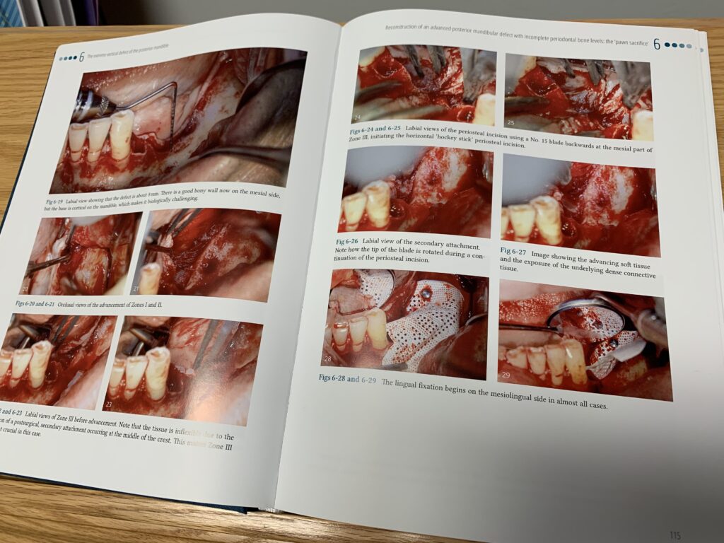
An entire section is dedicated to managing extreme vertical defects in the posterior mandible, including specific protocols to follow when there is scarred tissue or narrow basal bone. Dr Urban discusses the “pawn sacrifice,” or when you have to sacrifice a tooth to get your other chess pieces lined up—namely to facilitate membrane placement or allow more room for grafting. Other sections focus on anterior maxillary and mandibular vertical augmentation, soft tissue reconstruction in conjunction with bone grafting, reconstruction of the interimplant papilla—a particularly challenging thing to do—interproximal bone and soft tissue regeneration, and ultimate esthetics, among others.

What’s particularly engaging about this new book is that it doesn’t waste time or space with too much text that people won’t read. He knows that dentists are visual learners, so he steps back and lets the images tell the story. The book is a show-and-tell, so to speak. As Dr Urban writes in his preface, “The idea was to keep things as clear and simple as possible.”

He also includes “lessons learned” sections that give insight into what he learned from specific cases and how that knowledge can be applied to future clinical cases to improve precision and success. For example, in the chapter on the double strip graft, he shares that “free gingival grafts, and even a free CTG, result in a poorer esthetic outcome than a double SGG/collagen matrix combination.” Tips like this can go a long way in clinical practice, where the difference between success and failure can be as little as 1 mm of tissue.

Dr Urban describes it like this: “Parts of this book are like watching a surgical video with me, where I stop the video at the most important parts (sometimes frame by frame) and discuss with you, the reader, what I am thinking and doing at that step, and what my next step will be.” This is invaluable information for someone trying to learn from the best. And Dr Urban has proven himself to be among the best when it comes to ridge augmentation.

Vertical 2: The Next Level is a slam dunk. It’s easy to follow, full of stunning images, and can have significant impact on your clinical practice. The advance copies sold out within days. You’ll see why when you get your copy. Order today!


Vertical 2: The Next Level
Istvan Urban

This book is a continuation of the author’s best-selling Vertical and Horizontal Ridge Augmentation, but at a more advanced level. As such, readers should come armed with knowledge from the first book. In this volume the author delves deeper into the details of vertical ridge augmentation and really explores the intricacies of the procedures. Topics include treatment of the anterior maxilla, such as soft tissue reconstruction in conjunction with bone grafting as well as papilla reconstruction after bone regeneration, and the management of complications and difficulties in the posterior maxilla, such as deficient bony ridges and sinus involvement. Ridge grafting in the mandible is also included and given more detail than in the first book, this time focusing on larger defects and the different surgical steps required in native, fibrotic, and scarred tissue types around the mental nerve during flap advancement. The book features “lessons learned” sections that highlight learning objectives for improved precision, and the author demonstrates each technique step by step through meticulous case presentation, taking the time to explain his thought process and rationale every step of the way. In this must-have new publication, the procedures and techniques are kept simple, repeatable, and biologically sound so you can lower complication rates and improve predictability.

560 pp; 2,050 illus; ©2022; ISBN 978-1-78698-108-0 (BG143); US $280

Posted in Books, Implant Dentistry | Leave a comment

Fixed Prosthodontics: More Than Material(s) Selection

Reading time: 4 minutes

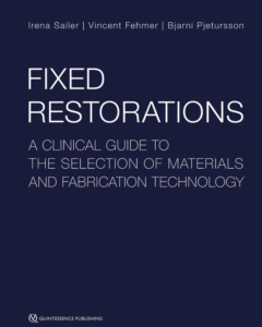
It’s no secret that technology is changing the way the world operates, and the field of fixed prosthodontics is no exception. With recent advances in CAD/CAM, 3D printing, and improvement in material properties, it can be difficult to stay up to date and informed on which treatment protocols are best. That’s why, when asked to write a foreword for Fixed Restorations: A Clinical Guide to the Selection of Materials and Fabrication Technology, Prof Dr Markus Hürzeler reconsidered his initial reaction—Do we need a book about fixed prosthodontics in this day and age?—because the answer is a resounding yes.

“It seems essential for the dental community to have an overview and guidelines of the current state of the art . . . considering the last decades of dentistry, it is apparent that a clinician will never be in the position of always being up to date in the fields of new materials and new manufacturing techniques of fixed restorations,” he summarizes. Additionally, while mechanistic therapeutic concepts and aspects of materials science have largely informed decision-making in restorative dentistry, these concepts have been challenged in recent years and replaced by ones that are biologically oriented. Periodontal, endodontic, and prosthetic approaches are all necessary for successful treatment outcomes with fixed prosthodontics. And, with the constant and rapid changes in technologies, laboratory technical expertise and familiarity with dental materials are also necessary.

Considering the different components needed to ensure successful prosthetic treatment, there was no better team to bring this book to life than Drs Irena Sailer, Vincent Fehmer, and Bjarni Pjetursson, who have united the fields of restorative dentistry, periodontology, and technical expertise. Staying up to date is much more manageable thanks to the trio’s efforts to collect the current evidence and best practices for material selection in fixed restorations. After all, “material selection” covers only part of the decision-making process—patient needs and fabrication technology are just as important as material properties when making these decisions.

Flowchart of material selection for tooth-supported single-unit restorations (minimally invasive, defect-oriented, and conventional).


Advances in technology mean that, in restorative dentistry, “the wax and modeling are evolving into software and mouse-clicks.” It’s true that technology has increased manufacturing efficiency, accuracy, and speed, but it has also allowed for improved patient communication. For instance, 3D-printed preparation guides or virtual mock-ups have proven to be useful tools for patient consultation as well as for the fabrication process. Likewise, augmented reality and facial recognition applications already exist for patient communication purposes, but show potential for use in fabrication technologies.

It might seem oxymoronic to compare one fabrication technology that could be used for communication to a communication technology that could be used for fabrication, but this sort of interplay couldn’t be more important for fixed prosthodontic treatment. After all, even when choosing a material, it is necessary to consider how a variety of clinical factors affect one another—do the quality and quantity of the remaining tooth substance allow for a conservative treatment like adhesive cementation, or are there factors at play that will require more preparation while remaining minimally invasive? Will any sacrifices, such as increasing the amount of preparation and material thickness to compensate for translucency, need to be made to ensure optimal esthetics? A single-tooth restoration is going to require different considerations than a multiple-unit restoration, so what kind of material and fabrication properties need to inform that treatment choice?

Translucency of different materials. From left to right; the leucite-reinforced glass-ceramic, feldspathic ceramic, zirconia, In-Ceram alumina, and In-Ceram zirconia samples have the same thickness (0.5 mm), and show obvious differences in translucency.


In addition to the sorts of questions a practitioner may pose to themselves, practitioners must also be ready to answer the patient’s questions, especially since it is so common for patients to research treatments themselves online before consulting a practitioner. It is important to respond to these informed questions and to correct any misconceptions if the patient’s information source is less than reliable, making it even more imperative to stay up to date with the current evidence, best treatment practices, and interplay of treatment considerations.

Many factors come together to ensure excellent fixed prosthesis treatment, and a whole book, split into four parts, was necessary to cover all of them. Fixed Restorations: A Clinical Guide to the Selection of Materials and Fabrication Technology was brought to life not only by the professional drive and expertise of its three authors, but also by their friendship, collaboration, and chemistry—ideals that can also be seen in good fixed prosthodontic treatment.


Fixed Restorations: A Clinical Guide to the Selection of Materials and Fabrication Technology
Irena Sailer, Vincent Fehmer, and Bjarni Pjetursson

In this timely and comprehensive guide to modern reconstructive dentistry, the authors showcase their expert knowledge of materials combined with innovative digital smile reconstruction technology to produce stunning results. The book is divided into four parts: basic information regarding materials and production processes, step-by-step clinical procedures with extensive case presentations, long-term outcomes, and management of complications. With over 2,000 clinical images and diagrams, backed up with the scientific evidence for recommendations, the best practice for tooth- and implant-supported fixed restorations is clearly described. Cases are presented in all different types of restorations, from minimally invasive veneers to implant-supported full-arch prostheses and everything in between. The vast clinical and technical knowledge and experience of the authors has resulted in a unique textbook that will aid in decision making regarding material selection and procedures for all patients in need of fixed restorations.

752 pp; 2,750 illus; ©2021; ISBN 978-1-78698-027-4 (B0274); US $360

Posted in Books, Implant Dentistry, Prosthodontics | Leave a comment

Visualizing the Dental Pulp

Reading time: 4 minutes

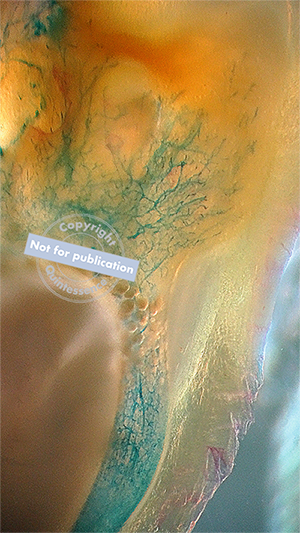
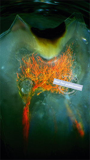
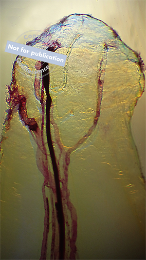
Craig Barrington, DDS, knows how to make teeth transparent. It seems implausible, but with enough know-how, you can diaphanize the hard enamel and dentin structures of extracted teeth to literally see what lies inside. It turns out, there is a lot to see.

“The dental pulp is an amazing and complex structure.” Dr Barrington continues, “There is not a lot of information available on the vascularity and nerve distribution within a human tooth. Most—if not all—pictures of the vascularity and innervation of the human pulp are artist-generated depictions of what they ‘believe’ or even ‘wish’ is inside our teeth. There is certainly more than we think hiding inside each and every single tooth.”

Like most people, I remember the first time I saw one of Dr Barrington’s images. The photograph showed the root of a tooth, but somehow, impossibly, the tooth structure was transparent, and the pulp canals were clearly visible. It was beautiful and arresting, and I had never seen anything like it. Even though I was familiar with his work, the collection of images in his new book, An Atlas of Dental Vascularity & Innervation, far surpasses anything I could have imagined. Throughout its pages, Dr Barrington shows us the beauty and grandeur of the dental pulp. The images speak for themselves, and they answer important questions about how nerves and blood vessels are distributed within teeth and the kinds of variation that exist for internal dental anatomy.

“We are missing a lot in dentistry. We extract and discard human teeth like they are trash. No other field of medicine trashes and discards human body parts at the same rate and with the same disregard as found in the field of dentistry. It would be my hope that the anatomical images featured in these pages would change this and set us on a new path.”

Preview Dr Barrington’s book here.


Craig Barrington, DDS, graduated summa cum laude in 1996 from the School of Dentistry at the University of Texas Health Science Center at San Antonio. Microscopes and histology have always been an interest of his, but the first time he looked at a transparent human tooth, he knew that this would be a lifelong focus of his research. To this end, Dr Barrington has made it his mission to understand every diaphanization method that he has come across. He has learned how, where, and why transparency can occur in a solid object. Dr Barrington holds a patent in histology and multiple copyrights. He maintains a private practice in Waxahachie, Texas.
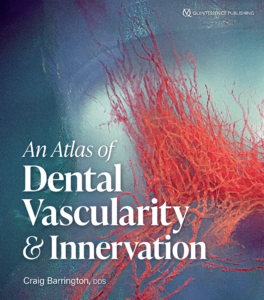
An Atlas of Human Dental Vascularity & Innervation
Craig Barrington

Our understanding of internal dental anatomy has remained limited by our inability to see inside a tooth without sectioning it. However, for one dentist working to find a way to see inside a tooth, the answer was diaphanization. This atlas represents the breathtaking results of photographing human teeth that have been made transparent. Dr Barrington has learned as many diaphanization methods as possible to understand how, where, and why transparency can occur in a solid object and translated that knowledge to transforming tooth structure. The images in this atlas showcase the internal anatomy of the teeth, with a special emphasis on the innervation and vascular structure and their distribution within the dentin chamber. For each image, the author follows a complex diaphanization method to make an extracted tooth transparent, before photographing the intact internal dental anatomy. Therefore, the images in this book display structures that have rarely been seen so clearly and in three dimensions, including the pulp chamber, apical anatomy, tooth channels, as well as pulpal pathology. This atlas pushes our understanding of internal dental anatomy and serves as an inspiration as to what one individual can do to advance knowledge within dentistry.

144 pp; 178 illus; ©2022; ISBN 978-1-64724-100-1 (B1001); US $104 (preorder)

Posted in Books, Dental Photography, Endodontics, Multidisciplinary | Leave a comment