February in Chicago: The Midwinter Meetings

Reading time: 2 minutes

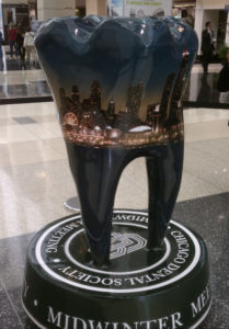
Every year at the end of February, the city of Chicago fills with thousands of the world’s top dental professionals who come to the city to attend one of many simultaneous meetings. For the Quintessence Publishing Chicago office, this week represents an annual opportunity to interact with the brightest minds of dentistry, meet with some of our authors, and represent the Quintessence brand at large.

For Quintessence the week revolves around the Chicago Dental Society’s Midwinter Meeting, taking place this year from February 23 to 25. The CDS Midwinter Meeting is one of the top 3 dental meetings in the United States and one of the top 10 conventions in the city of Chicago. Nearly 600 exhibitors participate, making this meeting one of the largest exhibits of dental products and manufacturers in North America.

Quintessence also attends Lab Day Chicago, hosted by LMT Communications February 24 and 25. LMT Lab Day brings over 250 exhibitors and 4,000 laboratory owners, managers, and technicians to the city. Just across the Chicago River, we also set up shop at the American Academy of Fixed Prosthodontics’s Annual Scientific Session February 24 and 25. This educational program brings together speakers from myriad scientific and clinical backgrounds to present concepts and clinical techniques based on scientific evidence and their own expert experience.

The Midwinter meetings are a busy, wonderful time in Chicago when the dentists outnumber the taxi cabs, and the three meetings that Quintessence attends are just a few of the many taking place in the city during this time. Midwinter has become a beloved tradition for the city, for the dental community, and for Quintessence.

Midwinter Meetings Quintessence Will Attend


CDS 152nd Midwinter Meeting: Booth 4408
Thursday through Saturday, February 23–25 in Chicago, Illinois

LMT Lab Day Chicago: Table A-12
Friday and Saturday, February 24–25 in Chicago, Illinois

AAFP 66th Annual Scientific Session: Navigating New Frontiers in ProsthodonticsBooths 122 & 123
Friday and Saturday, February 24–25 in Chicago, Illinois

Posted in Books, Esthetic Dentistry, Implant Dentistry, Multidisciplinary, Prosthodontics, Restorative Dentistry | Tagged , , , , , | Leave a comment

The Oral Microbiome and Why You Need to Know About It

Reading time: 7 minutes

Fusobacterium nucleatum, a bacterium commonly found in periodontal disease that has also been implicated in morbidities of other regions of the body, including colorectal cancer and adverse pregnancy complications such as preterm birth and fetal death. (Public Health Image Library image 2967, courtesy of the Centers for Disease Control and Prevention.)

When Wenche Borgnakke, DDS, MPH, PhD, was first asked to write a chapter in The Oral-Systemic Health Connection: A Guide to Patient Care (edited by Michael Glick, Quintessence, 2014) on the oral microbiome, she knew as much about it as most dental professionals do—next to nothing. “I told Michael, ‘I don’t know anything about the microbiome,'” she recalled. “And I didn’t! I only knew a little bit, like any other dentist or periodontist. I basically hunkered down the entire summer and read and read and read.”

The oral microbiome refers to the microoganisms in the oral cavity—bacteria, archaea, fungi, viruses, and protozoa. Recently developed technologies and computer capabilities now allow scientists to use DNA analysis to definitively “match” strains and subspecies of microbes from the oral cavity to those found elsewhere in the body. Using these data, scientists have been able to prove that microbes from the oral cavity can and do travel to other areas of the body. Now the big question for science is: What do they do when they get there?

A Delicate Balance

“The composition of the oral microbiome changes over time, and the microbiome for each person is different,” Dr Borgnakke explained. “Even the composition of biota in different locations of the same mouth is different, including around the same tooth. We usually talk about ‘periodontal pathogens.’ But, as a matter of fact, there are no studies that specifically identify any one or more bacteria that cause periodontal breakdown. So we have to get away from that notion that there are specific bacteria that cause gum disease and breakdown.”

Necrotizing ulcerative gingivitis, an opportunistic infection caused by an overgrowth of certain anaerobic microbes that flourish within deep periodontal pockets. (Used with permission from Oral and Maxillofacial Pathology: A Rationale for Diagnosis and Treatment, ed 2, by Robert E. Marx and Diane Stern.)

But where does moving away from this traditional view of bacteria in the oral cavity take us? Dr Borgnakke continued: “I spoke once with a Danish microbiology professor who came up with what was a completely brand-new idea for me: that the bugs in the mouth are actually there all the time. What he told me is that occasionally this homeostasis or balance is disrupted, either by antibiotics or by the host getting weak from stress or sick with something else, and then some bacteria will flourish in the more favorable living conditions while the numbers of other bacteria will decrease. So I maintain that periodontal disease is not really due to an infection—we usually consider infections to come from outside—but rather an overgrowth of bacteria that were already present but have taken over due to the homeostatic imbalance.”

The Traveling Oral Microbiome

During the research phase of her writing, Dr Borgnakke encountered a problem that many clinicians face when beginning to learn about the oral microbiome—it’s boring. She didn’t even want to read her own writing, so how could she make the information interesting enough to be accessible to others? Her solution: the metaphor of the traveling microbiome.

Helicobacter pylori can travel back and forth between the mouth and stomach and causes ulcers. (Courtesy of Dr David J. Kelly, University of Sheffield.)

“We used to think that stomach ulcers were caused by stress, then we realized they are caused by bacteria, such as Helicobacter pylori, that irritate the lining of the stomach. These bacteria are very difficult to kill and it can take a long time using many different antibiotics simultaneously. Over the course of treatment what can potentially happen is that via gastric reflux some of these bacteria can be regurgitated up in the mouth, which then serves as a reservoir. The bacteria can then live in the mouth and even re-infect the stomach.”

Traveling oral microbes have been identified all over the body, including the liver, the pancreas, the respiratory system, the gastrointestinal system, the synovial fluid of joints, and on the surfaces of intracardiac devices and other prostheses within the body. The microbes can travel to the respiratory system via aspiration, to the gastrointestinal system via swallowing and digestion, and to many other vital organs and areas of the body via the bloodstream.

Porphyromonas gingivalis is an anaerobic bacterium found in deep periodontal pockets that can enter the bloodstream and accumulate in atherosclerotic plaque. (Courtesy of Tsute Chen, PhD.)

“There are certain keystone bacteria that are usually present when there is periodontal breakdown,” Dr Borgnakke said, “and they are the ones that have developed really nasty mechanisms so they will not be discovered when they travel around the body. These bacteria can also have serious consequences when they travel outside the oral microbiome. For example, Porphyromonas gingivalis is often found in atherosclerotic plaque and can not only survive in the bloodstream but also multiply. We are getting closer and closer to proving that P gingivalis can destabilize these clots so that they loosen and have the potential to cause myocardial infarctions if they end up in the heart or ischemic strokes if they end up in the brain. There is another bacterium, Fusobacterium nucleatum, that is often seen in colorectal cancer lesions and also implicated in adverse pregnancy outcomes in the form of fetal death.”

What Next? Changing Our Habits Based on New Knowledge

The traveling oral microbiome. Microbes such as P gingivalis can enter the bloodstream and travel from the oral cavity throughout the circulatory system. (Adapted from At the Forefront: Illustrated Topics in Dental Research and Clinical Practice, edited by Hiromasa Yoshie.)

If the bacteria that can cause periodontal breakdown are always present but are only pathogenic when overgrowth occurs, how do patients and dental practitioners support this natural homeostasis? The answer may be to stop interfering.

“Stop using antibiotics!” Dr Borgnakke advised. “The guidelines say do not use antibiotics because they have been used frivolously and abused. Stop using antibiotics prophylactically, such as before cleaning or an extraction, because there is no evidence that it works. The guidelines have changed, but a lot of dentists will not change their habits. You should not give oral antibiotics to try to fight the bugs in the oral microbiome because they can’t penetrate the biofilm, which is where most of the bacteria we are concerned about reside. You kill good bugs along with the bad and skew the homeostatic balance. The antibiotics will kill some bacteria, causing others to flourish. And that imbalance is what people can die from.”

Another common practice that may need to be reevaluated is the use of mouthwash. Colloquially, mouthwash and other antimicrobial products (think hand sanitizer) are advertised to kill germs. But the oral microbiome contains both good and bad germs, and these products don’t differentiate. Similar to the use of antibiotics, the use of mouthwash can actually cause an overgrowth of disease-causing bacteria by upsetting the homeostatic balance of the microbiome.

What should we do then? Brush and floss. Tooth-brushing is beneficial to the gingiva not because of the ingredients in toothpaste but instead because of the manual biofilm removal that it facilitates. “The brushing is what is preventive for periodontal disease,” Dr Borgnakke explained. “You have to mechanically remove the biofilm.”

Resources for Further Education

With all the routes oral microbes have for traveling to other regions of the body and wreaking havoc, the stakes for dental professionals are high. Data regarding the oral microbiome and its effects on systemic health are fairly recent, and new research is being released at an alarming rate. It can be prohibitively time-consuming for clinicians to monitor every new development and determine how the new information should affect their practice. So how can clinicians and their teams keep up?
One option is to learn from Dr Borgnakke herself at The 2nd International Quintessence Symposium on Oral Health: The Oral-Systemic Health Connection, which will take place April 21–22, 2017 in Charlotte, North Carolina. This symposium brings together the most prominent clinicians and researchers on the subject of the oral-systemic health connection, including Dr Borgnakke. Dental professionals who attend can immerse themselves in 2 days of the most comprehensive and up-to-date knowledge on the associations between oral and systemic health.

“The oral microbiome is something all general dentists, dental hygienists, and periodontists should understand,” Dr Borgnakke stated. “I think it is a mystery that supposedly only a third of general dentists even have a periodontal probe in the examination kit. In other words they don’t routinely probe the space between the teeth and the gums. But we cannot neglect the basics. You absolutely must take care of the basics.”

The 2nd International Quintessence Symposium on Oral Health is the perfect opportunity for clinicans and their teams to improve their understanding of what goes on in the mouth at the microbial level so they can apply that knowledge to how they take care of both the basics and beyond.
Posted in Endodontics, Misc, Multidisciplinary, Periodontics, Research | Tagged , , , , , , , , | Leave a comment

Quintessence Roundup: February

Reading time: 6 minutes

Quintessence 2017 Catalog


New Titles in Books


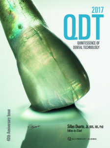
Quintessence of Dental Technology 2017
40th Anniversary edition!

Sillas Duarte, Jr (Editor)

QDT 2017 celebrates its 40th anniversary in style with beautifully presented original articles on new materials and techniques for achieving the utmost esthetic restorative results. This year’s focus is on challenging esthetic cases, treatment planning for various restorative options, new techniques using zirconia, and taking CAD/CAM beyond its limits. The State of the Art article presents the RAW digital workflow, and the Biomaterials Update features the Adhesive Restorative Complex concept. QDT’s new Masterclass article addresses the challenge of restoring a single central incisor. And the Masterpiece article this issue, presented by Naoki Hayashi, is a visual display of dental artistry.

256 pp; 1,000+ illus; ISBN 978-0-86715-736-9 (J0628); US $148


Vertical and Horizontal Ridge Augmentation: New Perspectives

István Urbán

This book expertly describes the key features of vertical and horizontal ridge augmentation, such as the surgical anatomy of the floor of the mouth, papilla reconstruction, the modified lingual flap and the preservation of the mental nerve during flap advancement, growth factor applications used in conjunction with perforated membranes, managing complications of membrane exposure and graft infections, and a step-by-step description of complete reconstruction of the severely resorbed maxillary ridge using GBR with simultaneous sinus augmentation. Almost all cases described include a retrospective section with suggestions on what could have been done differently to achieve even better results, making this book a frank, ambitious, and immensely informative text on the clinical practice of ridge augmentation with GBR.

400 pp; 1,229 illus; ISBN 978-1-78698-000-7 (B9096); US $244


Keep It Simple: Concept Porcelain Book

Paulo Battistella

In this book, the author expertly describes his techniques for layering restorative materials to more accurately recreate the esthetic qualities found in natural teeth. By separating the enamel layer into two distinct layers—enamel and enamel effects—the author achieves ideal biomimicry. Featuring expert photographs by Carlos Ayala, this book guides the reader through the refractory technique behind the author’s restorative creations.

236 pp; 588 illus; ISBN 978-85-7889-092-6 (B9986); US $150


The Workbook

Carlos A. Ayala Paz

With a reputation as one of dental photography’s most innovative experts, the author of this book elevates clinical photography to artistic expression. This workbook outlines the equipment, settings, and technical steps behind the author’s stunning photography and pairs his handwritten text with images of the production phases as well as the final photographic results. To see these photographs is to see the mouth in a way you can never forget, and this workbook will enable clinicians and lab technicians to likewise showcase the artistry of their own work.

252 pp; ISBN 978-85-7889-093-3 (B9987); US $220


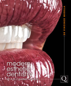
Modern Esthetic Dentistry: An A to Z Guided Workflow

Vincenzo Musella

This atlas synthesizes the latest technologic advances in esthetic dentistry and shows how to successfully integrate them into clinical practice. Part I begins with a chapter on dental photography that focuses on the correct settings and use of equipment needed to obtain high-quality images for documentation and communication with patients and the laboratory. Subsequent chapters present the steps involved in the esthetic preview, which provides the opportunity to try out a proposed treatment and make adjustments before undertaking any procedures, increasing patient acceptance and satisfaction. Part II presents clinical cases demonstrating the inverse layering technique for direct and indirect restorations, and Part III features clinical cases showcasing both traditional and alternative uses of lithium disilicate, an advanced restorative material with high esthetic potential and good strength. This is an excellent resource for clinicians looking to incorporate the latest techniques for predictable and stable esthetic results.

456 pp; 1,358 illus; ISBN 978-88-7492-038-9; US $191


New Issues in Journals


prd_banner

Featured article: Papilla Reformation at Single-Tooth Implant Sites Adjacent to Teeth with Severely Compromised Periodontal Support
Istvan A. Urban, Perry R. Klokkevold, and Henry H. Takei

Clinical Evaluation of the Influence of Connection Type and Restoration Height on the Reliability of Zirconia Abutments: A Retrospective Study on 965 Abutments with a Mean 6-Year Follow-Up
Giacomo Fabbri, Mauro Fradeani, Gianluca Dellificorelli, Marco De Lorenzi, Fernando Zarone, and Roberto Sorrentino

A Prospective Clinical and Radiographic Assessment of Platform-Switched Laser-Microchannel Implants Placed in Limited Interimplant Spaces
Myron Nevins, Sonia Leziy, Eric Kerr, Ulrich Janke, Giulio Rasperini, James Hanratty, Kirk Pasquinelli, Tiziano Testori, Cary A. Shapoff, and David M. Kim

ijp_banner

A Critical Review of Search Strategies Used in Recent Systematic Reviews Published in Selected Prosthodontic and Implant-Related Journals: Are Systematic Reviews Actually Systematic?
Danielle Layton

Sociodemographic, Educational, Behavioral, and Psychologic Factors Underlying Orofacial Esthetics and Self-Reported Oral Health
Cristina Gómez Polo and Javier Montero

Determining Favorable Maxillary Implant Locations Using Three-Dimensional Simulation Software and Computed Tomography Data
Tomoya Gonda, Koichiro Kamei, and Yoshinobu Maeda

jomi_banner

Thematic Abstract Review: Dental Implants in the Posterior Mandible: Complications and Management
Emad W. Estafanous

A Novel Osseous Densification Approach in Implant Osteotomy Preparation to Increase Biomechanical Primary Stability, Bone Mineral Density, and Bone-to-Implant Contact
Salah Huwais and Eric G. Meyer

Long-Term Results of Peri-implant Conditions in Periodontally Compromised Patients Following Lateral Bone Augmentation
Philip L. Keeve and Fouad Khoury

ebh_banner

Changing Our Long-Held Beliefs About Floss
Deborah M. Lyle

Quality Resources for Clinical Decision Making: Part 3. A Comparison Between Scaling and Root Planing and the Nd:YAG Laser in the Treatment of Periodontal Disease
Jane L. Forrest and JoAnn R. Gurenlian

Evidence on the Use of Probiotics in Periodontal Therapy
Silky Sharma

banner_ofph

Topical Review: Potential Use of Botulinum Toxin in the Management of Painful Posttraumatic Trigeminal Neuropathy
Nathan Moreau, Wisam Dieb, Vianney Descroix, Peter Svensson, Malin Ernberg, and Yves Boucher

Pain from Dental Implant Placement, Inflammatory Pulpitis Pain, and Neuropathic Pain Present Different Somatosensory Profiles
André Luís Porporatti, Leonardo Rigoldi Bonjardim, Juliana Stuginski-Barbosa, Estevam Augusto Bonfante, Yuri Martins Costa, and Paulo César Rodrigues Conti

Association Between Chronic Tension-Type Headache Coexistent with Chronic Temporomandibular Disorder Pain and Limitations in Physical and Emotional Functioning: A Case-Control Study
Rüdiger Emshoff, Felix Bertram, Dagmar Schnabl, and Iris Emshoff

Dental Meetings Quintessence Will Attend in February


ICOI Winter Symposium 2017: Solutions to Dental Implant Complications: Booth 101
hosted by the International Congress of Oral Implantologists, February 16–18 in New Orleans, Louisiana

CDS 152nd Midwinter Meeting: Booth 4408
hosted by the Chicago Dental Society, February 23–25 in Chicago, Illinois

LMT Lab Day Chicago
hosted by LMT Communications, February 24–25 in Chicago, Illinois

AAFP 66th Annual Scientific Session: Navigating New Frontiers in Prosthodontics
hosted by the American Academy of Fixed Prosthodontics, February 24–25 in Chicago, Illinois


Upcoming Quintessence Symposia


Posted in Books, Endodontics, Esthetic Dentistry, Implant Dentistry, International Journal of Evidence-Based Practice for the Dental Hygienist, Journal of Oral & Facial Pain and Headache, Journals, Misc, Multidisciplinary, Occlusion & TMD, Orthodontics, Pediatric Dentistry, Periodontics, Prosthodontics, Research, Restorative Dentistry, Roundup, The International Journal of Oral & Maxillofacial Implants, The International Journal of Periodontics & Restorative Dentistry, The International Journal of Prosthodontics, What's New | Tagged , , , , , , , , , , , , , , , , , , , , , , , , , , , , , , , , | Leave a comment

AUTHOR SPOTLIGHT: How Peter Sheridan Became Dentistry’s Expert on Clinical Photography, and Why You Should Be Doing What He’s Doing

Reading time: 6 minutes

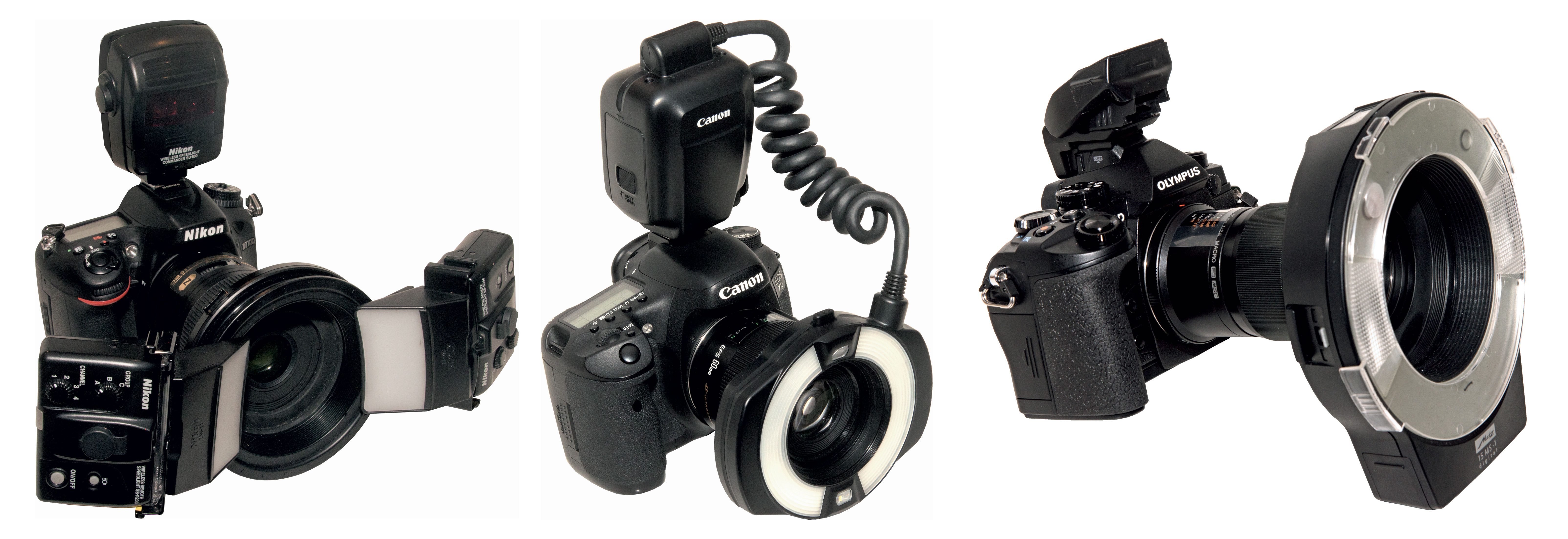
The three camera systems (Nikon, Canon, and Olympus) recommended by Dr Sheridan for clinical photography.

It is common practice in many dental clinics to perform a New Patient exam and create records that capture the exact status of the patient’s dental health. These records generally include radiography, a detailed patient history, and, in some practices, clinical photographs. Since the invention of digital cameras, clinical photography has become progressively more common in orthodontic and esthetic dentistry but not general dentistry. However, Australian dentist Peter Sheridan, AM, BDS, MDS, FICD, hopes to make clinical photography standard practice in every dental clinic.

“The standard assessment tools such as charting, clinical notes, and radiographs have not changed significantly in 40 years, although computerization has modified the process somewhat,” Dr Sheridan explains. “But the profession largely ignores the fact that none of these records show the mouth as it actually appears at the time of examination or treatment. From both a best-practice and a risk-management perspective, the use of routine clinical photography augments these other records and dramatically fortifies the dentist’s evaluation of the patient’s presenting condition and the treatment options. Photographs before, during, and after treatment can have enormous value in increasing patient understanding and acceptance of treatment.”

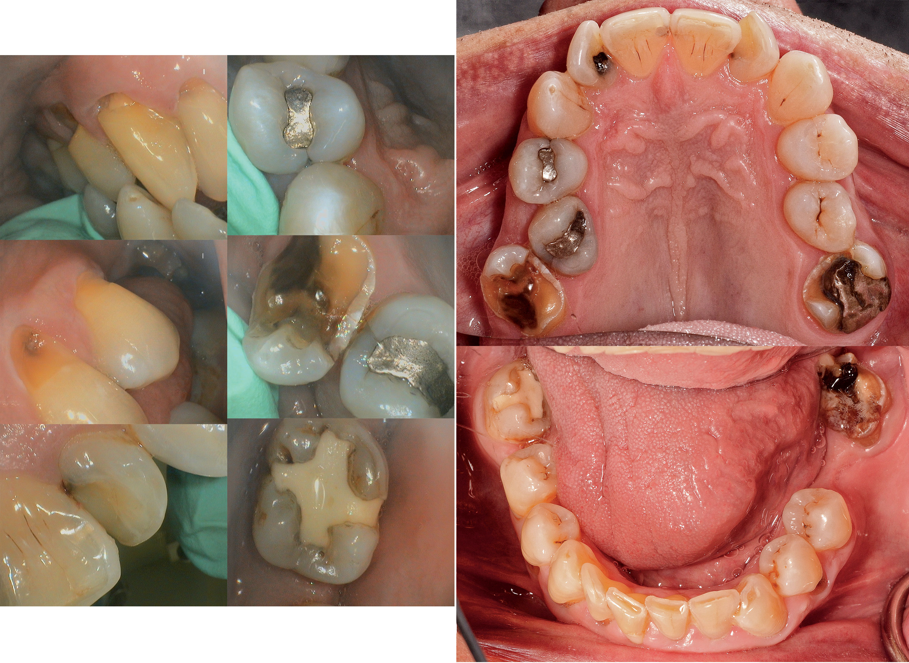
Images from an intraoral camera (left) versus images of the same patient using a DSLR camera (right).

Dr Sheridan’s interest in photography began long before his career in dentistry. When he was 12, he inherited the family’s Kodak Brownie camera after his father died. “There wasn’t much money for film,” he recalled, “but I practiced a lot and occasionally took photographs. I started my dental practice in 1971, and then I bought my first Nikon film camera and an 8 mm movie camera in the mid-1970s to record those magic moments of my kids growing up. I like instant gratification, so the delay between taking the photograph and receiving the finished prints or film a week or two later somewhat spoiled the photographic process for me. It was the excitement of digital photography and personal computers in the late 1990s that ticked all the boxes for me. I started to think that photography had a real place in dentistry as part of a digital suite of records.”

A self-taught photographer, Dr Sheridan’s crash course in photography came in 2005 when he decided to document the over 100 Art Deco radios he had been collecting as a personal hobby. Unable to find (or afford) a suitable professional photographer for the project, he decided to do the work himself and experimented with a barrage of technical photography equipment before settling on a simple and successful configuration: a camera, a bounce flash, and a tripod.

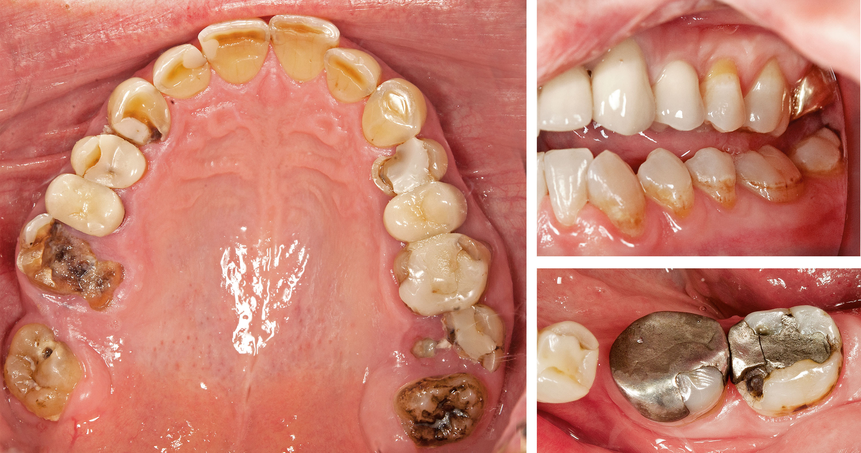
High-quality digital photographs can dramatically augment communication with patients regarding treatment concerns, especially in the posterior teeth.

“As I learned more about close-up photography,” Dr Sheridan said, “I realized that what I was doing in the surgery needed a complete update. At the time, most of the advice and teaching regarding clinical photography was based on outdated ideas from the film era and usually limited to anterior teeth. Our dental records are full of information, but there’s nothing that shows us exactly what we see in the mouth. I thought that clinical photography should add something significant to best practice in dentistry and also reflect reality. The images needed to be relevant and informational, of consistent high quality, and easily integrated into the workflow. I also felt that limiting clinical photography to the anterior teeth and cosmetic purposes was neglecting the bulk of everyday dentistry and taking the easy route. We are responsible for all of the oral tissues, both hard and soft, and even though photographing the posterior teeth is much harder, we need a system that allows us to comprehensively record the mouth as we see it.”

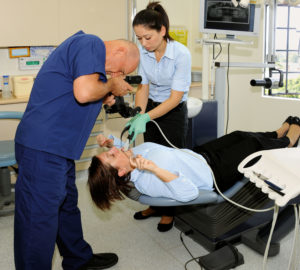
Dr Sheridan and members of his staff demonstrate how clinical photography involves collaboration between the photographer, an assistant, and the patient.

Dr Sheridan’s book, Clinical Photography in Dentistry: A New Perspective, outlines the system he has developed and is an accessible and comprehensive introduction to clinical photography for general dentists. The book covers the principles of clinical photography, recommended camera equipment and optimal settings, digital storage and management of the images, and standard views to be included in every patient’s record. The last chapter offers a structured approach to communicating with patients using the patient’s own clinical images, giving the dentist or hygienist an ethical and effective way to help patients make better decisions about their dental treatment. The book’s entry to the literature, which until now has lacked evidence of clinical photography’s value in general dentistry, is of significant importance.

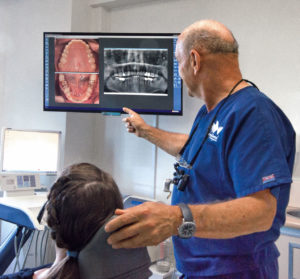
Digital photographs can be integrated into the same software as digital radiographs so that they can be viewed side-by-side when assessing the case and educating the patient.

“Dentistry is a profession with a strong commitment to change being supported by evidence-based research,” Dr Sheridan said. “Unfortunately, there is little published literature about clinical photography in dentistry. Until now there has been nothing to prove to a naysayer that there is an academic and common-sense foundation for the routine use of clinical photography in dentistry. My textbook is the first to identify and show the range of uses for clinical photography. The idea of increased acceptance of treatment plans is not just an assumed consequence of using photographs. My book is an attempt to bring a measure of academic rigor to this field in order to substantiate clinical photography’s value as an integral part of our recording and communication tools.”

Dr Sheridan hopes that his book will reach a whole new audience of dentists, hygienists, academics, and students all around the world, giving them a complete and rational perspective on how clinical photography is part of best practice in dentistry and how it can become one of their most valuable and informative tools.

Peter Sheridan, AM, BDS, MDS, FICD, is a dentist in Sydney, Australia, and has been in private practice since 1971. He is a clinical lecturer in the Faculty of Dentistry at the University of Sydney and presents courses, lectures, and master classes on clinical photography throughout Australia and New Zealand. His book, Dental Clinical Photography: A Guide to Standard Views (Bakelite, 2013) tailored the photographic protocol to general dentistry. He has a master’s degree in preventive dentistry and is a fellow of the International College of Dentists. Dr Sheridan is also an accredited professional photographer whose photographs and articles appear in specialist fine art journals, books, travel publications, museum catalogs, and print media. In addition, he is an internationally recognized historian, collector, and speaker on Art Deco design who has authored two photographic reference books on Art Deco radios, Radio Days: Australian Bakelite Radios (Bakelite, 2008) and Deco Radio: The Most Beautiful Radios Ever Made (Schiffer, 2014). In 2001 Dr Sheridan was awarded the Member of the Order of Australia by the Australian government for his international work in empowering people affected by multiple sclerosis.

He welcomes questions and comments and is available to help dental professionals incorporate clinical photograph into their practices. peter@clinicalphotography.com

Posted in Books, Esthetic Dentistry, Implant Dentistry, Misc, Multidisciplinary, Multimedia, Orthodontics, Pediatric Dentistry, Periodontics, Practice Management, Prosthodontics, Research, Restorative Dentistry | Tagged , , , , , , , , , , , | Leave a comment

In Memory of Robert Lee Vanarsdall, Jr, DDS

Reading time: 2 minutes

On January 1, 2017, the dental world lost a man of great force. Robert Lee Vanarsdall, Jr, DDS, leaves behind a superb legacy of contributions to orthodontic medicine. As longtime friends of Dr Vanarsdall, we at Quintessence recognize the void his passing leaves in our community as well as the educational foundation he has provided for the next generation of students and clinicians.

Dr Vanarsdall received his DDS from the Medical College of Virginia in 1970. Afterward he pursued specialty certificates in both orthodontics and periodontics at the University of Pennsylvania School of Dental Medicine. Dr Vanarsdall stayed on at UPenn as a professor, where he taught for more than 40 years. He also served as Chairman of the Department of Periodontics, Department of Pediatric Dentistry and directed the Orthodontics program from 1981 to 2011. In 2012, he was honored with the Dale B. Wade Award for Excellence in Teaching by the American Board of Orthodontics. At the time of his passing, he served as the Assistant Dean for Development for Dental Specialties and Chairman Emeritus, Department of Orthodontics at the University of Pennsylvania. In addition to his academic career, Dr Vanarsdall also maintained a private practice, where he continued to practice until his death.

The bibliography Dr Vanarsdall leaves behind is rich. He served as Editor-in-Chief for the International Journal of Adult Orthodontics and Orthognathic Surgery for 17 years in addition to other editorial board commitments. His relationship with Quintessence includes the development in 1980 of the Continuing Dental Education series, a collaboration between Quintessence and the University of Pennsylvania School of Dental Medicine, for which he authored Volume 2: Uprighting the Inclined Mandibular Molar in Preparation for Restorative Treatment. He coauthored the texts Applications of Orthodontic Mini Implants (Chicago: Quintessence, 2007) and Orthodontics: Current Principles and Techniques (St Louis: Mosby, 2017). He also published chapters in 12 other textbooks and over 100 scientific articles and abstracts. A worldwide lecturer, Dr Vanarsdall presented over 500 scientific lectures throughout his career.

Services were held January 5, 2017 at the St Thomas of Villanova Church on the Villanova University Campus in Villanova, Pennsylvania. In lieu of flowers, the family requests that donations be made to the Dr Robert Vanarsdall Fellowship Fund at the University of Pennsylvania School of Dental Medicine Department of Orthodontics.

Posted in Author Spotlight | Tagged | Leave a comment