Quintessence Roundup: September

Reading time: 5 minutes

New Titles in Books


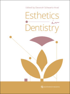
Esthetics in Dentistryschwartz-arad_cover

Devorah Schwartz-Arad

This book represents a unique collaboration between 17 internationally renowned female dentists as well as an insightful overview of contemporary esthetics. Topics include orthodontics and orthognathics, implants, restoration, rehabilitation, materials, trauma, and surgery. Each author also provides fascinating insight into her journey of success in a male-dominated industry.

352 pp; 812 illus; ISBN 978-1-85097-293-8 (B9090); US $198


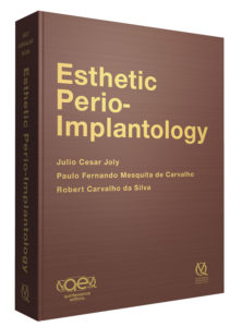
joly_coverAesthetic Perio-Implantology

Julio Cesar Joly, Paulo Fernando Mesquita de Carvalho, and Robert Carvalho da Silva

This book is the culmination of extensive research and experience in the field of esthetic mucogingival surgery and implant dentistry. The authors rely on clinical documentation and scientific evidence to establish innovative esthetic protocols for the management of mucogingival complications in implant dentistry. Each advanced surgical and restorative procedure is prefaced with the scientific research, historical background, and clinical experiences that led to its development by the authors. Facets of diagnosis and the many treatment options for each case are covered clearly with a strict evidence-based philosophy. The authors have streamlined their revolutionary techniques and innovations for standardized application, and each clinical case is presented step by step and in stunning photographic detail. This book will lay the foundation for the development of clinical skills that will lead to more esthetic outcomes in some of dentistry’s most challenging cases.

895 pp; 4,979 illus; ISBN 978-8-57889-086-5 (B9993); US $300


Dental Photography:chmielewski_cover
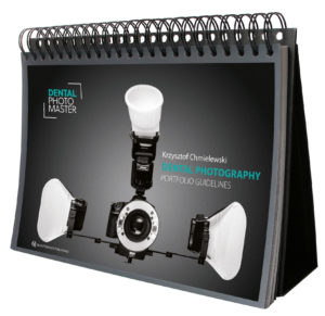
Portfolio Guidelines

Krzysztof Chmielewski

This practical atlas functions as a visual guide for using a camera in dental practice and achieving the essential photographic views. Individual views are detailed with recommended equipment setup, camera settings, necessary accessories, tips for the photographer, and instructions for the patient. The book is designed to function as a stand-up display, providing ease of use to the photographer when used chairside. Whether used as a refresher by an experienced dental photographer or as a guide for instructing staff members on dental photography protocols, this atlas is sure to become a mainstay in clinical practice.

59 pp; 64 illus; ISBN 978-1-85097-297-6 (B9094); US $98
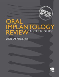
 

Oral Implantology Review: A Study GuideNEW iBOOK!
Oral Implantology Review: A Study Guide

Louie Al-Faraje

This comprehensive examination workbook provides more than 700 practice questions on oral implantology. Topics include medical problems, biomedical sciences, radiology and computer-assisted technology, anatomy, biomechanics, patient data, treatment planning, principles of implantology, bone and soft tissue grafting, implant prosthodontics and occlusion, esthetics, maintenance, pharmacology, and complications.

232 pp; 74 illus; ISBN 978-0-86715-721-5 (B7215); US $108


New Issues in Journals


prd_banner

Featured article: A Classification System for Peri-implant Diseases and Conditions

Hector L. Sarmiento, Michael R. Norton, and Joseph P. Fiorellini

Comparison of Marginal Bone Changes with Internal Conus and External Hexagon Design Implant Systems: A Prospective, Randomized Study

Lyndon F. Cooper, Dennis Tarnow, Stuart Froum, John Moriarty, and Ingeborg J. De Kok

Transmucosal Implant Placement with Submarginal Connective Tissue Graft in Area of Shallow Buccal Bone Dehiscence: A Three-Year Follow-Up Case Series

Martina Stefanini, Pietro Felice, Claudio Mazzotti, Matteo Marzadori, Enrico F. Gherlone, and Giovanni Zucchelli

 ijp_banner

Editorial: The Digital Revolution’s Impact on Prosthodontics

Carlo Marinello

A 5-Year Retrospective Assay of Implant Treatments and Complications in Private Practice: The Restorative Complications of Single and Short-Span Implant-Supported Fixed Prostheses

Jason Hsuan-Yu Wang, Roy Judge, and Denise Bailey

Effect of Double Screw on Abutment Screw Loosening in Single-Implant Prostheses

Young-Gun Shin, So-Yun Kim, Ho-Kyu Lee, Chang-Mo Jeong, So-Hyoun Lee, and Jung-Bo Huh

jomi_banner

Thematic Abstract Review: The Issue with Tissue: How to Treat Peri-implantitis

Martin Osswald

Prevalence of Interproximal Open Contacts Between Single-Implant Restorations and Adjacent Teeth

Spyridon Varthis, Anthony Randi, and Dennis P. Tarnow


New Titles in Multimedia


Cell-to-Cell Communication:celltocell_oralhealth_cover
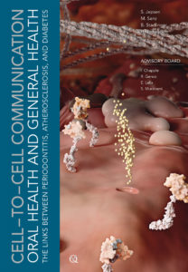
Oral Health and General Health—The Links Between Periodontitis, Atherosclerosis, and Diabetes

Søren Jepsen, Mariano Sanz, Bernd Stadlinger, and Hendrik Terheyden

Can periodontitis or other inflammatory processes of the oral cavity contribute to systemic conditions such as atherosclerosis or diabetes? Can they negatively influence their course? This new installment in the Cell-to-Cell Communication series addresses these issues by focusing on the associations linking periodontal and systemic health. The film visualizes the highly complex intercellular interactions and signaling pathways. Periodontal pathogens are invasive and can spread throughout the body via the bloodstream. Locally produced pro-inflammatory mediators can also shed into the circulation. This new film shows the dissemination of bacteria in periodontitis, the impact of periodontitis on the cardiovascular system (atherosclerosis), the effect of periodontitis on the metabolism (type 2 diabetes), and the effect of dental treatment.

17 minutes; DVD-ROM; ISBN 978-1-85097-288-4; US $128


Dental Meetings Quintessence Will Attend in September


CDA Presents: The Art and Science of Dentistry: Booth 1604

hosted by the California Dental Association, September 8–10 in San Francisco, California


2016 FACD Annual Scientific Session & Trade Show: Booth 200

hosted by the Florida Academy of Cosmetic Dentistry, September 8–10 in Orlando, Florida


AAP 102nd Annual Meeting: Booth 1433

hosted by the American Academy of Periodontology, September 10–13 in San Diego, California


SCAD 2016: Extravaganza

hosted by the Society for Color and Appearance in Dentistry, September 15–17 in Chicago, Illinois


AAOMS 98th Annual Meeting: Scientific Sesions & Exhibition: Booth 712

hosted by the American Association of Oral and Maxillofacial Surgeons, September 21–23 in Las Vegas, Nevada


ICCMO 27th Annual Bernard Jankelson Memorial Lecture Forum: Multidisciplinary Approach To Optimal Care

hosted by the International College of Craniomandibular Orthopedics, September 22–24 in Scottsdale, Arizona


Spear Faculty Club Summit

hosted by Spear Education, September 29–October 1 in Scottsdale, Arizona

Posted in Books, Esthetic Dentistry, Implant Dentistry, Journals, Misc, Multidisciplinary, Multimedia, Periodontics, Prosthodontics, Research, Restorative Dentistry, The International Journal of Oral & Maxillofacial Implants, The International Journal of Periodontics & Restorative Dentistry, The International Journal of Prosthodontics, What's New | Tagged , , , , , , , , , , , , , , , | Leave a comment

Accessing the Collections of the World’s Largest Museum

Reading time: 6 minutes

Fetal skulls

The Smithsonian Institution in the United States has long been a top destination for tourists, but it is also a valuable resource for researchers. It houses over 138 million objects across 19 museums and galleries in 2 cities. In addition to the publicly exhibited works, the Smithsonian also maintains over 150,000 cubic feet of archives. It was in these archives that Lawrence Freilich, DDS, PhD, found the unique collection that would inspire his first book.

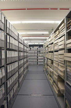
Records storage at the Smithsonian Archives. Could one of these boxes hold a 100-year-old fetal skeleton?

Records storage at the Smithsonian Archives. Could one of these boxes hold a 100-year-old fetal skeleton?

“I had retired from teaching at Georgetown University, so I was looking for a way to get involved with some research pertaining to what I had done while at Georgetown, which was on bone regeneration,” Dr Freilich recollected. “I was familiar with the Smithsonian and beginning to get an interest in anthropology, so I called and asked if they had anyone I could get in touch with. They connected me with Dr David Hunt, who is an expert in forensic anthropology.”

David Hunt, PhD, D-ABFA, is the Assistant Collections Manager for the Smithsonian’s Department of Anthropology at the National Museum of Natural History (NMNH) and oversees all of their human skeletal collections. “My job is to facilitate outside research by connecting researchers to the collections that match their study goals, scheduling appointments for access, and answering any questions they may have,” Dr Hunt said. “We have a study going on right now that involves decorative filing of the teeth during the Mississippian era and another on the burial practices of Chinese immigrants working in the Alaskan salmon canneries in the late-1800s. Typically I have 65–70, sometimes over 100, external researchers who come over the course of the year and usually during the summer.”

Dr Freilich was one of those researchers who floated in without a clear idea of what he was looking for. “I asked if [Dr Hunt] had any specific projects I could get involved with, and he said there was a European anthropologist who had come in and started working on the collection of fetal skeletons they had. While I was looking at some of those skeletons I started picking up jaw bones and just became fascinated. I couldn’t recall any literature that displayed fetal jaw bones close-up and in detail, so we started putting together this study.”

That study became the Atlas of Fetal Jaw Development published this year. As explained in the first chapter of the book, the NMNH’s fetal collection was largely donated by doctors and academics so its specimens are exceptionally well documented in terms of age, gender, and ethnic origin. Dr Hunt described the history of this collection and how original collector Aleš Hrdlička obtained them as one of the most interesting parts of the study.

“Just to see and understand the breadth of how Hrdlička had the medical community involved and aware of this collection was impressive. Of course, [Franklin Paine] Mall and [Daniel Smith] Lamb were the two main contributors, but Hrdlička also obtained individuals from dozens of other obstetricians and gynecologists in the DC and Baltimore areas. The social milieu of today versus the early 1900s is different, so we no longer receive similar donations en masse. I think there are differences in the attitudes of how bodies are turned over for scientific studies than there were back in the turn of the century. There are numerous body laws that came about in the 1950s and ‘60s where unclaimed bodies are no longer the property of the state. They’re still the responsibility of the state, but unless there is paperwork explicitly giving permission to donate the body to science then the state must bury or cremate the body.”

The fetal collection involved in Dr Freilich’s study was, according to Dr Hunt, almost completely derived from pathological death or spontaneous, rather than elective, abortion. When a similar event occurs today, the family and the hospital must fill out paperwork through a body donation program in order to send the fetal remains to an institution like the Smithsonian. While the body laws are necessary to protect an individual’s right to bodily autonomy, the red tape has caused a shortage in cadavers available to the scientific community, making the NMNH’s fetal collection all the more impressive.

“The anatomical collections,” Dr Hunt said, “can serve as a template for archaeological study since we know who they were and what they died of. We have researchers come in to measure bone metrics from our anatomical collections to assist in determining the age and sex of other skeletal remains.”

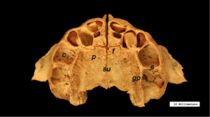
Occlusal view of the articulated maxillae in a later-stage fetus with teeth beginning to form in the dental crypts.

Occlusal view of the articulated maxillae in a later-stage fetus with teeth beginning to form. c, crypts; f, incisive fissure; gp, greater palatine nerves and vessels; p, palatine process; s, interdental septae; su, midline suture.

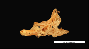
Buccal view of the right maxilla. Note the sponge-like surface of the bone.

Buccal view of the right maxilla. Note the sponge-like surface of the bone. Anteroposterior length: 15.3 mm. b, body; f, frontal process; i, infraorbital foramen; ig, infraorbital groove; z, zygomatic process.

Anatomical collections are also useful for developmental studies, which is what piqued Dr Freilich’s interest. “The bone that becomes the tooth socket isn’t even present at 2–3 months of fetal age. So in the middle-stage fetuses—that’s 3-4 months in utero—you can observe the tooth sockets beginning to develop. Just the architecture and the surface anatomy of the jaw bones were fascinating to observe under the magnification of the camera lens. The surface structure of these bones appears sponge-like and displays an abundance of neurovascular channels. As the bones mature, those channels gradually diminish in size and are largely replaced by a more dense, sturdy bone tissue. This is necessary since the jaw bones and other skeletal elements need much more strength following birth.”

“What surprised me the most,” Dr Freilich said, “was how the skeletons were preserved and stored. They are all disarticulated as in fetal life and are stored in boxes. The entire skeleton is in one container. So it took some time to sort through the collection and find enough maxillae and mandibles in good shape from each age group to photograph.”

But photographing fetal bones isn’t easy. “You can put a thigh or an arm bone on a table and take a good picture very easily because they lie flat. But for the jaw bones we had to use pans of sand to angle the bones perfectly and keep them steady. We used tweezers to handle the bones because they are so fragile. When we were arranging the full skeletons in the pans of sand, the hardest part was the vertebrae. This is because in fetal life, each vertebral bone remains in three separate pieces, which are not joined yet. It was truly fascinating to see such tiny bones and handle them.”

Buccal view of the right mandible. Pans of sand were used to stabilize the bones for photography. During post-production of the images, the sand was removed and markers were added to show scale and significant structures.

Buccal view of the right mandible. Pans of sand were used to stabilize the bones for photography. During postproduction of the images, the sand was removed and markers were added to show scale and significant structures. Anteroposterior length: 20.6 mm. b, body; c, condyle; co, coronoid process; m, mental foramen; n, mandibular (sigmoid) notch; r, ramus.

For researchers interested in collaborating with the Smithsonian, Dr Hunt’s advice is to visit the website or call the manager of the collection you’re interested in. Appointments typically run 2–4 months ahead for scheduling, especially during the summer months.

For Dr Freilich, he simply sent in his curriculum vitae and the Department of Anthropology staff went about “matchmaking.” The result, Atlas of Fetal Jaw Development, is both a fascinating study on fetal jaw development and a testament to what discoveries open access in the research sector can foster.


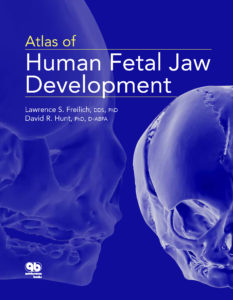
Atlas of Human Fetal Jaw Development

Lawrence Freilich and David Hunt

This atlas presents a study of human fetal jaw development with a primary focus on the hard tissues of the maxilla and mandible. High-definition photographs are featured throughout of the well-preserved upper and lower jaw elements from the human fetal skeletons contained in the collections of the Smithsonian National Museum of Natural History.

72 pp; 83 illus; ISBN 978-0-86715-716-1 (I7161); US $37.99; iBook only

Posted in Books, Misc, Multidisciplinary, Pediatric Dentistry, Research | Tagged , , , , , , , , , , , , , , , | Leave a comment

Quintessence Roundup: August

Reading time: 3 minutes

Books


New Titles

Oral Implantology Review: A Study GuideOral Implantology Review: A Study Guide

by Louie Al-Faraje

This comprehensive examination workbook provides more than 700 practice questions on oral implantology. Topics include medical problems, biomedical sciences, radiology and computer-assisted technology, anatomy, biomechanics, patient data, treatment planning, principles of implantology, bone and soft tissue grafting, implant prosthodontics and occlusion, esthetics, maintenance, pharmacology, and complications.

ISBN: 978-0-86715-721-5; 232 pp; 74 illus (softcover); USD $108


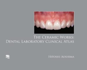
The Ceramic Works: Dental Laboratory Clinical AtlasThe Ceramic Works: Dental Laboratory Clinical Atlas

by Hitoshi Aoshima

Written by a master ceramist with a keen eye for the essence of color and form, this atlas features 60 clinical cases, an appendix of tooth samples in ceramic, and commentary with case details and enlightening laboratory tips for achieving optimal ceramic restorations. The author discusses the development of his internal live stain technique and shares many insights about subtle differences that are key to making restorations look natural. A beautiful reference on ceramic restoration for dental technicians.

ISBN: 978-0-86715-709-3; 104 pp; 361 illus; USD $82


Implants in the Esthetic Zone: A Step-by-StepGrunder_Cvr Treatment Strategy

by Ueli Grunder

With a focus on oral surgery and prosthodontic techniques, this book presents effective implant treatment strategies for the esthetic restoration of anterior teeth. In addition to reviewing biologic principles, treatment planning, indications, esthetic analysis, and prosthetic options, the author details key considerations in each stage of treatment, including gentle tooth extraction, precise implant positioning, criteria for one- and two-stage implant placement, and most importantly, a wide range of soft tissue management techniques to prevent or compensate for tissue loss in the esthetic zone. With ample cases presented in a step-by-step fashion, this book is a useful resource that can help clinicians avoid failures and achieve optimal esthetic success.

ISBN: 978-1-85097-283-9; 848 pp; 4,049 illus; USD $348


Journals


New Issues

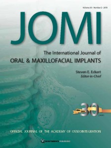
The International Journal of Oral & Maxillofacial ImplantsThe International Journal of Oral & Maxillofacial Implants

Editor-in-Chief: Steven E. Eckert

Thematic Abstract Review: Do We Get Improved Precision of Implant Fixed Dental Prostheses with Digital Scanners and Computer-Aided Design/Computer-Assisted Manufacture?

Jan-Eirik Ellingsen

Effect of Preseating, Screw Access Opening, and Vent Holes on Extrusion of Excess Cement at the Crown-Abutment Margin and Associated Tensile Force for Cement-Retained Implant Restorations

Rodrigo A. Jimenez,Tatiana Vargas-Koudriavtsev

Long-Term Esthetic Evaluation of the Roll Flap Technique in the Implant Rehabilitation of Patients with Agenesis of Maxillary Lateral Incisors: 10-Year Follow-Up

Ioannis K. Konstantinidis, Konstantinos D. Siormpas, Eleni Kontsiotou-Siormpa, Miltadis E. Mitsias, Georgios A. Kotsakis


Journal of Oral & Facial Pain and HeadacheJournal of Oral & Facial Pain and Headache

Editor-in-Chief: Barry J. Sessle

The Effectiveness of Physiotherapy in the Management of Temporomandibular Disorders: A Systematic Review and Meta-analysis

Maria Paço, Bárbara Peleteiro, Jos. Duarte, Teresa Pinho

Systematic Review and Meta-analysis of the Efficacy of Oral Medications Compared with Placebo Treatment in the Management of Postherpetic Neuralgia

Sabrina Salah, Lesly Thomas, Saravanan Ram, Glenn T. Clark, Reyes Enciso

Trigeminal Neuralgia Induced by Sour and Spicy Foods: What Is the Underlying Mechanism? A Case Report

Elon Eisenberg


Dental Meetings Quintessence Will Attend in August


The Quality of Esthetics: Booth E3

hosted by Ivoclar Vivadent, August 19–20 in Mexico City, Mexico

Posted in Books, Esthetic Dentistry, Implant Dentistry, Journal of Oral & Facial Pain and Headache, Journals, Prosthodontics, Restorative Dentistry, Roundup, The International Journal of Oral & Maxillofacial Implants, What's New | Tagged , , , , , , , , , , , , , , , , , , , , , | Leave a comment

Remembering a Legend: Charles J. Burstone

Reading time: 3 minutes

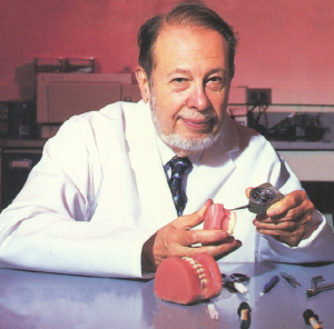
On February 11, 2015, Dr Charles J. Burstone, orthodontist, educator, and researcher, passed away. The following is the Tribute and In Memoriam published in Dr Charles J. Burstone’s final book project titled The Biomechanical Foundation of Clinical Orthodontics (Quintessence Publishing, 2015). Dr Burstone coauthored the book with Kwangchul Choy, DDS, MS, PhD. The Tribute was written by Dr Choy and the In Memoriam was written by Michael R. Marcotte, DDS, MSD. To find out more information about The Biomechanical Foundation of Clinical Orthodontics or to order a copy, visit www.quintpub.com.


Screen Shot 2015-05-12 at 2.45.29 PM

Dr Charles J. Burstone

(1928-2015)

Tribute

Sadly for us, after finishing this book, a giant fell.

Most of the contents of this book are based on Dr Burstone’s energetic and rigorous research for more than 200 research articles. The format of this book was adopted from the lectures on biomechanics that we gave at the University of Connecticut and Yonsei University for many years. Over the last 3 years, my work with Dr Burstone to convert
those lectures and ideas into this book was one of the most challenging, most exciting, and the happiest moments in my life. As one of his students, an old friend, and a colleague, I have to confess that all of the concepts in this book are his.

In the beginning, Force was created with the Big Bang. Fifteen billion years later, Newton discovered the Law of Force in the universe. However, the knowledge of how to control orthodontic force remained an occult practice that was only revealed through years of orthodontic apprenticeship. It was Dr Burstone who uncovered the magic
and found the principles governing this treatment method that was once thought to be mysterious. There is no doubt that the Law of Orthodontic Force was his discovery.

I would like to share Dr Burstone’s words from his last lecture with me on February 11, 2015, in Seoul: “Don’t believe blindly in experience, but believe in theory, and think creatively.”

“My father shaped my body; you shaped my thoughts.” Charles, our dearest friend, may you rest in peace.

                                                                                                                                                                                          -Kwangchul Choy, DDS, MS, PhD

In Memoriam

Dr Charles J. Burstone, orthodontist, educator, researcher, and friend to many, passed away February 11, 2015, of an apparent heart attack in Seoul, Korea. He died doing what he loved to do and in a place where he loved to be.

Dr Burstone is well known for the development of the field of scientific biomechanics. He was a master teacher in orthodontics who could bridge the gap between understanding key engineering concepts and applying them to clinical practice. He made biomechanics understandable by showing how to use simple engineering principles to solve most orthodontic problems. His Segmented Arch Technique was developed through the use of sound engineering principles.

Dr Burstone was unwavering in his enthusiasm for student learning and was dedicated to ensuring clinical excellence in his students. When I was a student, I can remember reviewing a patient’s treatment plan and him asking me, “What do you want to do with the lower incisors and why?” He emphasized the importance of having clear, specific, and defensible treatment objectives and then designing mechanical plans that would achieve those treatment objectives, step by step.

Over his lifetime, Dr Burstone trained hundreds of orthodontists first at Indiana University and then later at the University of Connecticut. He served as Department Chairman while at each institution. He was a recipient of many awards and honors and remained active in organized dentistry throughout his life, serving in many positions and lecturing around the world.

Dr Burstone also had a deep connection to Korea. He served there during the Korean War, and his photographs and movies from this period depicted everyday Korean life in a time of conflict. The National Folk Museum in Seoul developed an exhibit around his images entitled “Korea, 1952,” and his images were also used in a Korean documentary about the Korean War. He was devoted to Korea, and it is indeed fitting that his last lecture was delivered in Seoul.

He truly loved his profession and was a beloved mentor and colleague to many. He leaves the worldwide orthodontic community to mourn his passing.

-Michael R. Marcotte, DDS, MSD

Posted in Books, Misc, Orthodontics | Tagged , , , , | Leave a comment

JOMI Celebrates 30 Years

Reading time: 4 minutes

JOMI 2_cover

The following is the editorial from the March/April 2015 issue of The International Journal of Oral & Maxillofacial Implants (JOMI), which was written by JOMI Editor-in-Chief Steven E. Eckert, DDS, MS.

Happy Birthday JOMI!

This journal began publication 30 years ago with two issues in its first year. Editor Emeritus William R. Laney’s first editorial announced the launch of the journal, and his second editorial commented on a newspaper article in which Ann Landers, the columnist, responded to a reader who submitted a question on dental implants. Ann Landers consulted with a “prominent dental school dean” who described dental implants as providing 5 years of support for dental prostheses. At that time, the 5-year expectation was based upon the Harvard/NIH consensus conference on dental implants. Also at that time, osseointegration had been described but was not yet well appreciated. Dr. Laney’s editorial described the need for better understanding of dental implants, which demands a clear appreciation of the science behind the implants that clinicians use in treatment of their patients. The time when implant performance could be summarized in one sentence had passed; the era of modern, data-based implant performance had arrived.

With this editorial, Dr. Laney identified the future of JOMI. This is not a journal with a 30,000-foot-high overview; instead, it is a journal that assesses implant dentistry at the microstructure level. The journal was created to identify scientific research and translational clinical applications derived from these investigations. The journal began with clear intent, and I am sure you will agree that it has lived up to this vision.

Today, the journal is published every other month along with additional “supplemental” issues that appear almost every year. The supplements tend to be focused on a specific theme, while the regularly scheduled publications provide research under the topics of “Implant Science” and “Clinical Applications.” These bimonthly issues of the journal present the breadth and the depth of the primary investigations that are ongoing throughout the world. In addition, the reader benefits from secondary research that combines information from primary research to assimilate (synthesize) information into a more compelling appreciation of the topic at hand. The secondary research is presented in the form of systematic reviews with meta-analyses, which then lead to clinical recommendations derived directly from the data or indirectly from consensus interpretations of the data.

In its 30 years, JOMI has laid the groundwork for general acceptance of the concept of osseointegration throughout dentistry. The first decade of the journal provided glimpses into the future with the promise of improved implant survival for all bone types, shortened healing periods, identification of less invasive grafting techniques and materials, better definition of loading protocols, and myriad of other scientific and technical advances.

Today, we utilize technology that we might not have envisioned three decades ago. Three-dimensional imaging is almost ubiquitous. Affordability of cone beam computerized tomography has made this technology available to most clinicians. Not only can we now visualize the body in three dimensions, but we also are able to make use of this knowledge to create surgical guides and to provide real-time surgical guidance. No matter where we travel, dental meetings are not complete without some discussion of implant dentistry. The dental implant provider industry has grown remarkably during the same timeframe.

With this much progress in a 30-year span, we probably should start to wonder where the next 30 years will take us. Will true biologic substitutes be available at an affordable price in the year 2045? Do we envision a point where a mixture of cells—today, we would be thinking of stem cells, but the future may change this—is delivered to a deficient anatomical area, allowing the tissues that were missing from that area to regrow? Maybe the description of a “mixture of cells” might be too limiting; perhaps the answer to the consequences of tooth and tissue loss will come in some form that we are not even considering today. Of course, the development of a method of intervention that will re- create anatomical structures is valuable only if patients can maintain the re-created structures. This statement circles back to the notion that patients must be committed to the maintenance of the technically ingenious devices that are provided to them. When a third dentition is established, it would be gratifying to know that traditional dental disease will not befall that third dentition.

When I entered dental school 41 years ago, the dean stood up and told the students that we had chosen the wrong career path. His statement was based upon the notion that a dental caries vaccine and a periodontal disease vaccine were on the near horizon. His statement was somewhat apologetic because we had all worked very hard to gain admission to dental school and his discouraging news seemed to make dentistry a dead-end profession.

Today, we continue to wait for these vaccines. As we wait, we should probably gain an appreciation that when we can prevent dental disease, we have actually done exactly what the goal of dentistry has always been, and that is to make dental care delivery unnecessary.

This is somewhat akin to what has happened in a few different areas of medicine. An example that comes to mind is the near total elimination of polio as a disease entity. I imagine that when the polio vaccine was developed, there were some physicians who feared that their careers would end when polio disappeared. Although there may have been a few physicians who were so entrenched in the treatment of this disease that its virtual eradication did indeed eliminate the need for those physicians, most of those polio specialists simply transitioned to a different area of expertise.

When the cure comes to dentistry, as it surely will at some point, perhaps before JOMI reaches 60, I hope that dentistry will celebrate this success. When this journal is not needed, that means that the battle against edentulism will have been won. Won’t that be great?

© 2015 BY QUINTESSENCE PUBLISHING CO, INC.

JOMI is published by Quintessence Publishing and is the official journal of the Academy of Osseointegration. For more information about JOMI, visit quintpub.com.

Posted in Implant Dentistry, The International Journal of Oral & Maxillofacial Implants | Tagged , , | Leave a comment