Quintessence Authors Share Their Scary Cases

Reading time: 5 minutes

If you’ve worked in the medical field, you’ve had a day in the clinic that was scary, for one reason or another. And just like in horror movies, there are different kinds of scary. There are experiences that shock you, gross you out, or leave you with a sense of impending doom. You might even receive an unexpected phone call. In the spirit of Halloween, we asked Quintessence authors to share their scary cases with us. If you read this article after dark, you might want to keep the lights on.

Cranial Fracture

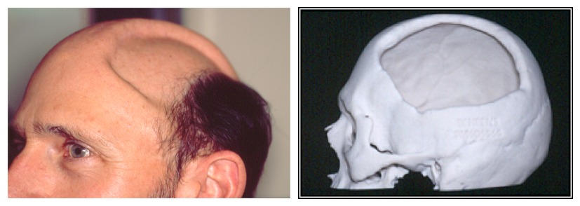
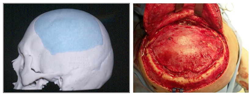
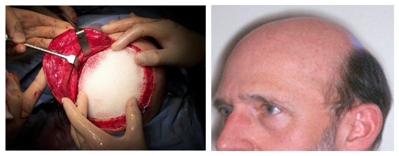
Our first case is from Dr Johan Reyneke, author of three editions of Essentials of Orthognathic Surgery. This patient, a 46-year-old man who was a dentist himself, sustained a cranial fracture after a cycling accident. The fracture developed an infection, resulting in sequestration of the outer table of his cranium. On examination, the inner cranial bone was found to be mobile, putting the brain in jeopardy. Dr Reyneke was able to correct the defect by using a 3D computer-generated model and creating a patient-matched implant. He used a unilateral coronal approach to place the implant, resulting in a full recovery and renewed confidence for the patient.

Hepatic Carcinoma Metastasis

Dr Paul Coceancig remembers a bloody case from his first day of work: “I was in Singapore General Hospital in the oral surgery clinic. It was my first day on the job. I was called by a very anxious dental nurse, who pulled me into the dental office of my new and very pale junior colleague who had an 80-year-old—and extremely pale—patient with literally a liter of bright red blood around his boots. My colleague had just done a mucosal biopsy of what turned out to be a hepatic carcinoma metastasis that had lodged inside the cheek as a bright red blister. They apparently bleed without any chance of control, but that’s not something anyone knew at the time.

Without really spending much time to think about it, I jumped right in with a huge stitch that curved around the mandible to compress the facial artery that was supplying the area in question. My buddy was patting me on the back, shaking my hand, and the old bloke was starting to pink up. But then my senior colleague—the one who ran the clinic—walked in, saw all the blood, and cut the stitch that was tied over the skin as it wasn’t ‘intraoral.’ I didn’t have time to say ‘NOOOOO!’

The bleeding immediately restarted, and nothing she did seemed to work, until she too asked for the same stitch to be retied, so I stepped right back in.

We all learned something that day.”

Severe Dental Trauma

Dr David Sarver looks back on a challenging case of a patient who experienced severe dental trauma and serious bodily injury, which is also featured in his new book Dentofacial Esthetics: From Macro to Micro: “Emergencies don’t often happen with orthodontics, but this one rattled me a bit. This 24-year-old woman was referred to me after being struck by a car while living abroad. She had recovered from her fractured limbs and ribs, and now it was time to take care of her teeth, which had been stabilized with a bonded fiber strip.

It was hard to see what I was looking at with all the acrylic and gingival hypertrophy (Fig 1), so I took a CBCT scan to see what was under there. Most of the tooth crowns and roots were intact, but some were avulsed and/or fractured (Fig 2). My first thought was that all of these traumatized teeth would have to be extracted, because saving them would certainly be impossible. But then I considered the bony defect and cascade of effects that would follow, a fate I wanted to avoid for this young woman. Instead, my goal was to preserve as much bone as possible. It was worth a try to save whatever I could.

“The story certainly didn’t end here, but the scary part was over.”—Dr Sarver

After administering local anesthesia, I gently removed the bonded splint and all the bonding material, and then I used a diode laser to remove all the hypertrophic gingiva (Fig 3). Full fixed appliances were placed, and within 6 weeks the maxillary anterior teeth were almost in contact with the mandibular incisors, and the CBCT showed intact roots that, surprisingly, were asymptomatic (Fig 4). No teeth were lost on my watch, and the patient had the stability she needed to continue restorative treatment. The story certainly didn’t end here, but the scary part was over.”


Hereditary Gingival Fibromatosis

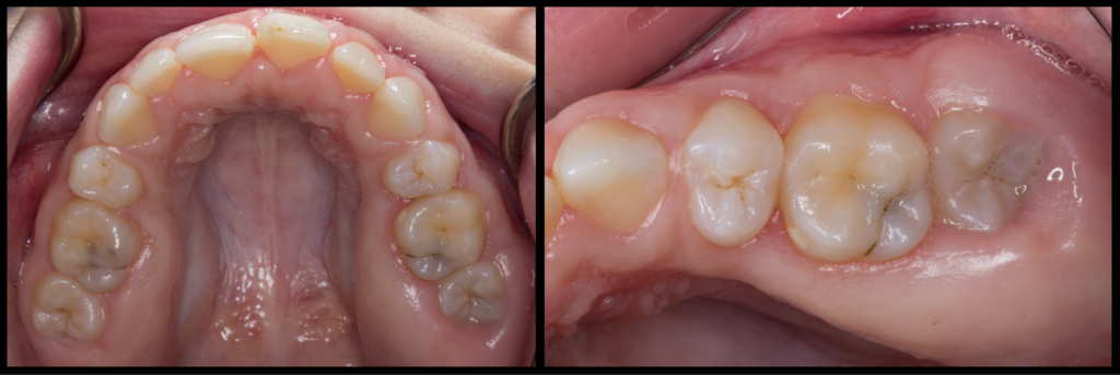
Dr Gustavo Avila-Ortiz sent us some before and after photos of this case of hereditary gingival fibromatosis, a very rare periodontal condition treated with gingivectomy. Very scary for someone who’s never encountered it before!

Vampire Dentist

Dr Daniel Ninan, author of Dentistry and the Pregnant Patient, recalls an otherworldly experience: “When I was working as a mobile dentist, I went to a facility to see a patient. The patient was very nice. However, when I entered the patient’s room, the other person in the room held up a cross and kept the cross between me and them. I wondered if she perhaps thought I was a vampire.”

Face-Eating Incident

Remember the face-eating incident in Miami in 2012? Yoh Sawatari, DDS, author of Surgical Management of Maxillofacial Fractures, was the facial trauma attending who got the initial call that day, and to this day he remembers the call he received from the intern: “Sawatari, I have a patient who had his face eaten by another person, what do you want me to do?” As he was contemplating his career choices, he got a call from the director of trauma: “Plastics wants to take the case, do you mind?” Dr Sawatari didn’t mind at all.


Thank you to the Quintessence authors who shared their scary cases with us! Be sure to check out their books to learn more about how to treat cases, whether they’re scary, routine, or somewhere in between. Happy Halloween!

Posted in Misc | Leave a comment

Now Is the Time to Embrace Dental Photography in Your Practice

Reading time: 7 minutes

Written by: Bryn Grisham

In the last decade, dental photography has gone from being an elite hobby for true photographic enthusiasts to a best practice and an essential part of daily record keeping. It wasn’t that long ago that photography was considered a demanding and expensive pastime that offered minimal clinical benefit. Certainly dental photography was always useful for communication and documentation, but it wasn’t practical for the average clinician. Even when the cost was not a deterrent, clinicians still had to contend with the steep learning curve; it takes a lot to understand the basics of photography and to master the concepts and equipment. All in all, it didn’t seem like a good investment. However, most of that has changed, and it is easy to see why.

Cost and Accessibility

Gone are the days of expensive and complicated equipment that is only available from professional photography shops.

Gone are the days of expensive and complicated equipment that is only available from professional photography shops. Everything you need to take photos in clinical practice has become more widely available and more affordable. And digital photographs are now cheap and easy to store. What this means is that the biggest financial hurdles have been lowered, considerably.

Clinical Context

It has never been easier to document cases and communicate using digital photographs. Regardless, the question that dentists kept coming back to is whether dental photography could really make a difference in their daily practice, especially when it seems like a time-consuming process. Everyone has heard how digital photography can improve patient communication, but can it really increase patient acceptance? With the rise of fast and easy digital mockups, old notions have been challenged and now we have a more tangible incentive for using photography in clinical practice every day to plan and discuss treatment plans with patients.

Simple and Effective Protocols

Even if the costs are reasonable and more clinicians want to use dental photography in their practice, that doesn’t mean there aren’t any hurdles left to clear. Learning the basics of dental photography is still a real obstacle.

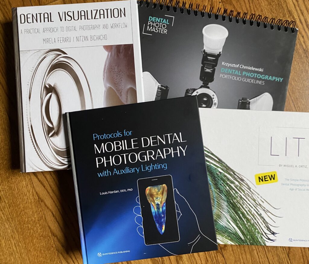
This is where Quintessence comes in. Our expert authors have figured this stuff out and are sharing what they know to help you. We have multiple best-selling textbooks on dental photography that meet you wherever you are—whether you are looking for a traditional textbook to start at the beginning, a fun new clinical approach that makes it easy, a quick reference for staff members, or a how-to for taking clinical photos with a mobile phone. We’ve got you covered!

Comprehensive Textbook

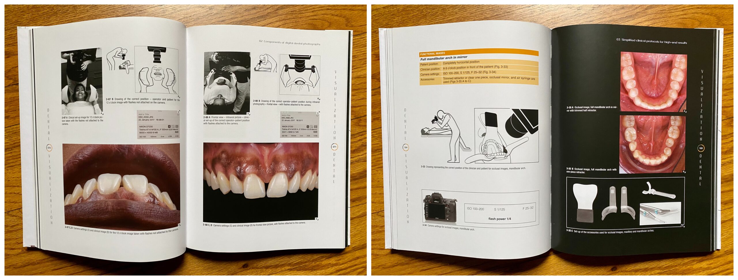
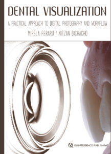
Our most traditional textbook is Dental Visualization: A Practical Approach to Digital Photography and Workflow. Drs Feraru and Bichachu cover everything from photographic principles to products to troubleshooting, as well as laying out how any specialist can apply this knowledge into the daily workflow of their practice. It has everything you need to know.

Best Clinical Manual

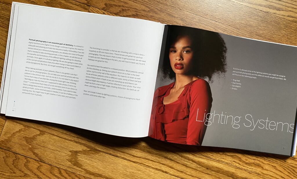
In 2019, we launched our best-selling book by Dr Miguel Ortiz, Lit: The Simple Protocol for Dental Photography in the Age of Social Media. Dr Ortiz breaks down photographic concepts to their simplest components, which helps readers gain confidence and start using a DSLR camera in practice right away. This book takes the mystery out of photography and will help anyone find their footing. Moreover, the author’s enthusiasm for the topic comes through from the beginning and makes dental photography seem very manageable.

Quick Guide

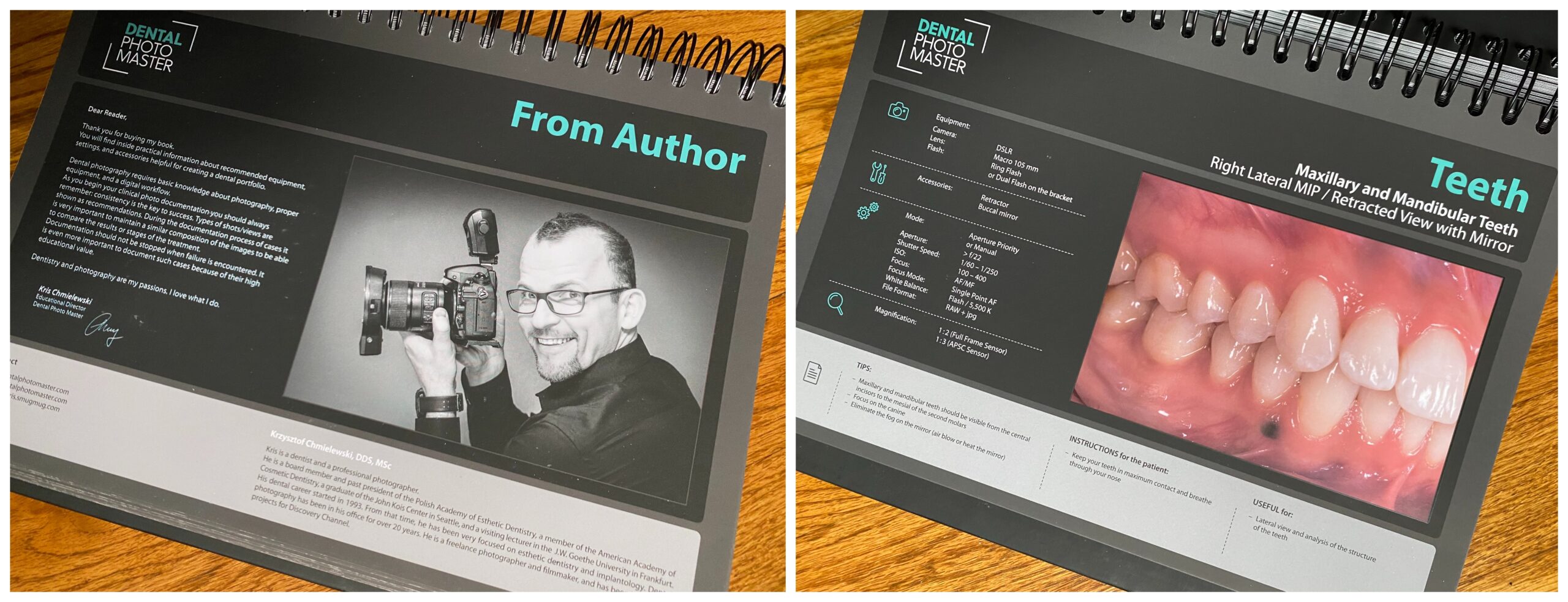
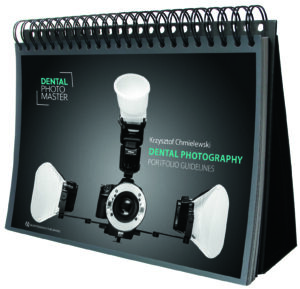
Dr Chmielewski has written the essential quick guide, Dental Photography: Portfolio Guidelines. The brilliance of his shot-by-shot guide is that it outlines the equipment, accessories, mode, magnification, and tips for every clinical photograph that you would need to take. This is a go-to reference for you and your staff.

Dental Photography with Your Phone

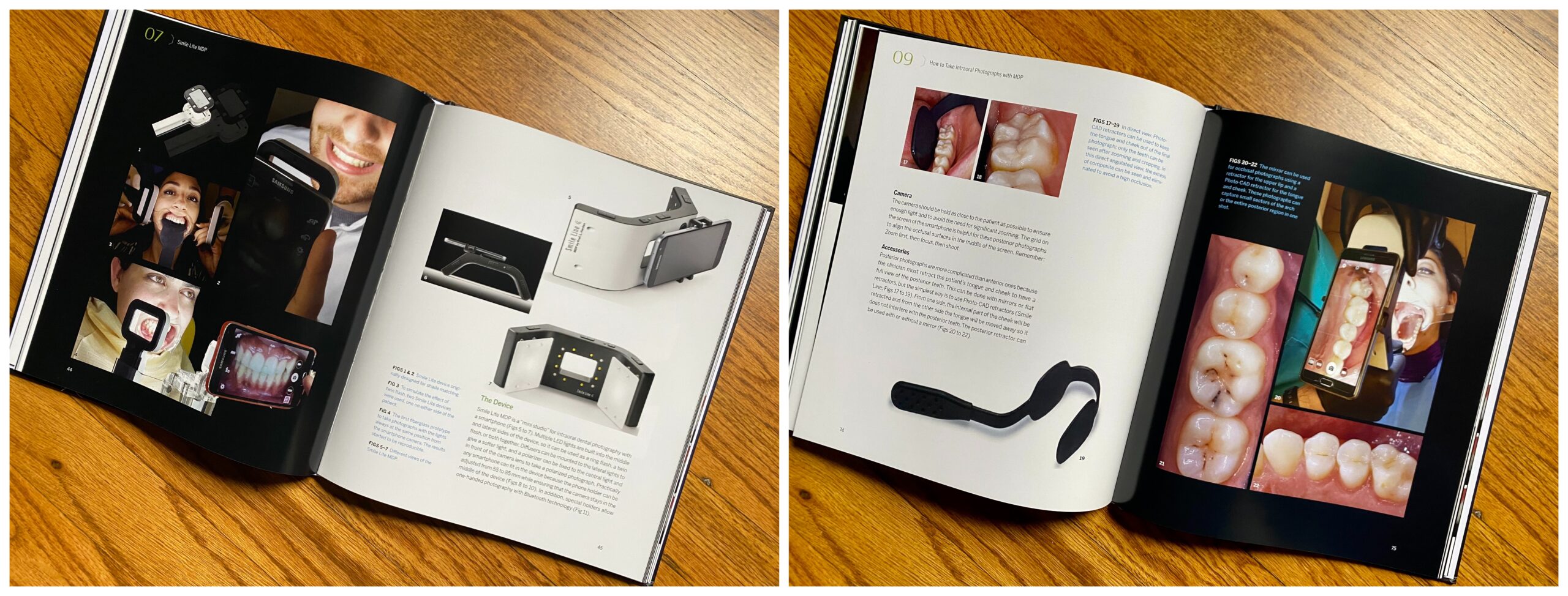
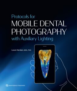
Finally, our newest book on dental photography is for every clinician who wants nothing to do with using a DSLR camera in their practice (or who wants more convenience and flexibility). Dr Hardan shows you how to use the camera on your phone with a few simple accessories to achieve outstanding dental images. His book, Protocols for Mobile Dental Photography with Auxiliary Lighting, makes it clear that you really can get similar-quality images with a smartphone camera as with a DSLR camera; you just need to know how to use it.

Dental photography is not going anywhere, and sooner or later, most clinicians are going to have to find a way to make it work in their practice. Make certain that you have the best information on hand to help you make that transition a success.

Dental photography is not going anywhere, and sooner or later, most clinicians are going to have to find a way to make it work in their practice.


Dental Visualization: A Practical Approach to Digital Photography and Workflow
Mirela Feraru and Nitzan Bichacho

This book provides the practicing clinician with a clear and concise guide to dental photography and its role in modern dentistry. The main aspects are broken down into easy-to-follow chapters, all lavishly illustrated with detailed images. Aspects such as the importance of digital documentation, camera components, and photographic equipment as well as simplified protocols for high-end results, different dental specialties, and troubleshooting are all covered within the book’s pages. The authors have condensed their extensive knowledge and expertise into a book that will surely prove invaluable to all those working within modern dental practices.

248 pp; 556 illus; ©2018; ISBN 978-1-78698-004-5 (BG128); US $158

Lit: The Simple Protocol for Dental Photography in the Age of Social Media
Miguel A. Ortiz

In the age of digital dentistry, dental providers are under increased pressure to demonstrate proficiency in dental photography for the purposes of documentation, shade matching, and laboratory communication. Expertise in this area is fast becoming part of the standard of care and also has added value for clinicians who are looking to market themselves online. This book is geared toward practitioners who want to master dental photography and build their social media presence. Written for visual learners, this book breaks down the fundamentals of dental photography by outlining the key concepts, equipment, and lighting as well as by introducing “The Simple Protocol”—the basic day-to-day intraoral protocol that shows how easily clinical photography can be incorporated into the clinical workflow. That is where most photography books end, but this author also explores advanced techniques and demonstrates how to achieve some of the most characteristic looks in artistic dental photography, including the glossy effect, chiaroscuro, chromaticity, and texture manipulation, as well as a simple setup for taking photographs in the dental laboratory. Finally, the author provides fresh insight into the ever-changing world of digital marketing and explains what you need to know to reach your market on social media.

248 pp; 357 illus; ©2019; ISBN 978-0-86715-802-1 (B8021); US $148

Dental Photography: Portfolio Guidelines
Krzysztof Chmielewski

This practical atlas functions as a visual guide for using a camera in dental practice and achieving the essential photographic views. Individual views are detailed with recommended equipment setup, camera settings, necessary accessories, tips for the photographer, and instructions for the patient. The book is designed to function as a stand-up display, providing ease of use to the photographer when used chairside. Whether used as a refresher by an experienced dental photographer or as a guide for instructing staff members on dental photography protocols, this atlas is sure to become a mainstay in clinical practice.

59 pp (spiral bound with slipcase); 64 illus; ©2016; ISBN 978-1-85097-297-6 (BG120); US $98

Protocols for Mobile Dental Photography with Auxiliary Lighting
Louis Hardan

Billions of photographs are taken worldwide each day, and most of them are captured with a smartphone. In the context of clinical dentistry, many clinicians use their smartphone to take clinical photographs for communication or documentation simply because they cannot operate or afford a professional camera. Author Louis Hardan understands too well how a poorly taken photograph can compromise the quality of dentistry delivered to the patient, which is why he launched the Mobile Dental Photography (MDP) project in 2012. He wanted to find a way to make documentation and communication easy and affordable to all dentists and laboratory technicians, including students. In this book he outlines how to use the smartphone camera with auxiliary lighting and accessories to take photographs for treatment planning, documentation and self-evaluation, communication with the patient and laboratory technician, tracking the evolution of treatment, as well as for publishing, lecturing, and insurance and medicolegal purposes. He also shows how to take artistic photographs to wow your followers on social media and effectively market your practice online. You really can get similar-quality images with a smartphone camera as with a DSLR camera; you just need to know how to use it. This book is the key.

128 pp; 315 illus; ©2020; ISBN 978-0-86715-946-2 (B9462); US $75

Posted in Books, Dental Photography, Multidisciplinary, What's New | Leave a comment

High-Quality Photography at Your Fingertips (or in Your Pocket)

Reading time: 5 minutes

Written by: Leah Huffman

Clinical photography can be an intimidating thing for a dentist. Everyone knows you need to document your cases, but do you need to invest in an expensive professional camera setup and learn all the ins and outs to achieve the level of quality you need? Absolutely not. Dr Louis Hardan has an alternative that is easier, cheaper, and certainly more convenient.

After all, with good lighting, a smartphone camera is perfectly able and sufficient to take a good-quality photograph.  

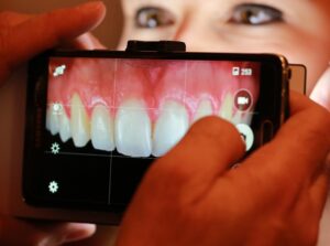
In 2012 he started the Mobile Dental Photography (MDP) project in hopes of finding a way to make documentation and communication easy and affordable to all dentists and laboratory technicians, including students. Partnering with Smile Line, he created a lighting device that can be attached to your smartphone camera to ensure good-quality photographs. Dr Hardan recognized that many dentists were already using their smartphone cameras anyway, so why not turn that very capable device into a full studio for excellent photography? After all, with good lighting, a smartphone camera is perfectly able and sufficient to take a good-quality photograph. As Dr Hardan explains it, “Since smartphone cameras are so popular and evolving very quickly during the last few years…I decided to offer a practical, affordable, and easy solution to dentists by developing MDP.” And thus the Smile Lite MDP device was born.

“In truth, we make a good image; we don’t take it.”—Louis Hardan  

However, Dr Hardan recognizes that you can’t just slap the Smile Lite on a phone and start taking excellent photographs. First you need to understand how to take clinical photographs properly and how the smartphone camera works. He explains that “In truth, we make a good image; we don’t take it.” So before clinicians can start making good images, they need to understand the basics, which is why he wrote his recent book, Protocols for Mobile Dental Photography with Auxiliary Lighting, to be released in October 2020.

In this book he outlines how to use the smartphone camera with auxiliary lighting and accessories to take photographs for treatment planning, documentation and self-evaluation, communication with the patient and laboratory technician, tracking the evolution of treatment, as well as for publishing, lecturing, and insurance and medicolegal purposes. He also shows how to take artistic photographs to wow your followers on social media and effectively market your practice online.

Dr Hardan wants dentists to be excited about MDP for its many advantages: “Every dentist has a free camera in his pocket with high performance. Since photography is a good management of light, MDP will offer to the dentist a mini studio where he has all kinds of light used in professional photography, from the ring flash and lateral flashes to diffused and polarized light. Since the light is continuous, the user can see on his screen the final result before taking the picture.” Better yet is that this system costs way less than a professional-grade DSLR camera and is way more intuitive to use.

“Every dentist has a free camera in his pocket with high performance. Since photography is a good management of light, MDP will offer to the dentist a mini studio where he has all kinds of light used in professional photography.”—Louis Hardan  

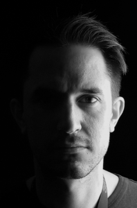
One of the coolest things about MDP is that you can use it to take artistic photographs like half-illuminated portraits, birefringence photographs, and glam shots of lips and teeth. Dr Hardan shows in his book how to take all of these with your smartphone. Social media has quickly become an effective marketing tool for dentists, and shots like these can give you the edge you need to succeed. As Dr Hardan underscores, “Lots of pictures on social media are low quality because of the lighting used. High-quality photographs with MDP can be used to market our work. If the dental treatment is perfect but the pictures are bad in quality, the marketing message will be very bad. That is why the dental pictures should be perfect, and by adding artistic pictures, the impact on social media will be very high.”

If you’re already using a DSLR system you love, don’t change a thing. But if you’re struggling with your existing system, aren’t taking the types of images you’d like to, or simply aren’t taking any images at all, consider what MDP can do for you and your practice. Dr Hardan sums it up like this: “MDP in daily use is very practical, easy, lightweight, can be disinfected, and helps to take all kinds of photographs.” You simply cannot go wrong with MDP for clinical photography.

To preview Dr Hardan’s new book, click here.


Protocols for Mobile Dental Photography with Auxiliary Lighting
Louis Hardan

Billions of photographs are taken worldwide each day, and most of them are captured with a smartphone. In the context of clinical dentistry, many clinicians use their smartphone to take clinical photographs for communication or documentation simply because they cannot operate or afford a professional camera. Author Louis Hardan understands too well how a poorly taken photograph can compromise the quality of dentistry delivered to the patient, which is why he launched the Mobile Dental Photography (MDP) project in 2012. He wanted to find a way to make documentation and communication easy and affordable to all dentists and laboratory technicians, including students. In this book he outlines how to use the smartphone camera with auxiliary lighting and accessories to take photographs for treatment planning, documentation and self-evaluation, communication with the patient and laboratory technician, tracking the evolution of treatment, as well as for publishing, lecturing, and insurance and medicolegal purposes. He also shows how to take artistic photographs to wow your followers on social media and effectively market your practice online. You really can get similar-quality images with a smartphone camera as with a DSLR camera; you just need to know how to use it. This book is the key.

128 pp; 315 illus; ISBN 978-0-86715-946-2 (B9462); US $75

Posted in Books, Dental Photography, Multidisciplinary, What's New | Leave a comment

Is Microneedling Right for Your Practice?

Reading time: 9 minutes

Written by: Marieke Zaffron

With the rise of platforms like Instagram and all the photos of young, beautiful people—edited or not—that come with it, it’s no surprise that demand for cosmetic treatments is at an all-time high, with no sign of slowing down. Fortunately, the technologic developments that enable increased exposure to sometimes unachievable beauty standards have been accompanied by technologic developments in the fields of esthetics and dermatology, making treatments to combat aging safer than ever before.

Now you—and your patients—may be asking, what do skin treatments have to do with dental treatment? Why are dentists offering things like Botox? Shouldn’t that be left to dermatologists?

The fact is that dentists are remarkably well-equipped to deal with procedures affecting the skin of the face. Many of the lines and wrinkles that patients most want to minimize occur around the mouth, and who has a better understanding of the anatomy in this area than dentists? Dentists are very familiar with administering anesthesia, and many have already used adjunctive products like platelet-rich plasma (PRP), platelet-rich fibrin (PRF), and hyaluronic acid (HA) for other applications.

Dentists are remarkably well-equipped to deal with procedures affecting the skin of the face.

When we perform prosthetic treatment planning, we consider the face as a whole. It’s not just the teeth, but the smile. It’s not just the smile, but the face. Asking patients what they would like to change about the esthetics of their smile provides an opportunity to discuss the face as a whole. Patients may be unaware that things such as wrinkles, scars (including acne scars), and more can be treatable at the dentist’s office, often for a relatively low cost, with a low risk of complications, and with great success.

Microneedling in particular, which is discussed in both Drs Catherine Davies and Richard Miron’s PRF in Facial Esthetics and Drs Arun Garg and Renato Rossi’s upcoming Dermal Fillers for Dental Professionals, has many advantages compared with other treatments. As Drs Garg and Rossi explain, “Dermal microneedling restores firmness and elasticity to the face. It has become a popular alternative to more invasive procedures such as laser skin resurfacing and deep chemical peeling for skin rejuvenation. It is noninvasive and does not damage the skin, and it can be performed using topical anesthesia, another feature that is attractive to patients.” Drs Davies and Miron add that the healing time for this method is shorter than other options (only 1 to 2 days), is appropriate for all skin types, and is well tolerated by patients. The procedure is also quick to perform in office—15 to 20 minutes—and is easy to master.

How Microneedling Works
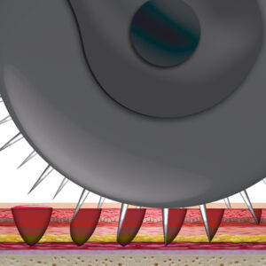
Microneedling is a minimally invasive treatment that involves using a specialized tool such as a roller or a pen to make tiny puncture wounds on the surface of the skin—approximately 200 punctures per square centimeter, according to Drs Garg and Rossi. Because it is technically an injury, the body responds with wound-healing processes, including the release of growth factors. As Drs Davies and Miron explain, “Microneedling induces soft tissue fibroblast proliferation and collagen and extracellular matrix deposition, re-epithelialization, and angiogenesis. … It has also been shown to stimulate collagen production by creating microchannels into the dermis and initiating a repair process.” Importantly, scars will not form as a result of microneedling. The injury is controlled and minimal, just enough for neocollagenesis to produce new collagen and elastin to tighten the skin and improve its architecture.

Who Can Benefit

Microneedling began as a treatment for wrinkles, but it now has multiple indications, including wrinkles, stretch marks, hair loss, hyperpigmentation, ultraviolet damage, acne, and scars (including acne scars). There are, however, some contraindications to microneedling. Microneedling has been shown to be less effective on ice pick scars than other types of atrophic scars. Patients taking blood thinners should not undergo microneedling therapy because of an increased risk of bleeding. (Though the procedure ordinarily results in very minimal bleeding.) Diseases of the skin such as eczema, rosacea, and malignancies are further contraindications.

However, most patients who have undergone microneedling have been very happy with the results. Studies have shown microneedling to be very effective in the treatment of wrinkles as well as most types of atrophic scars, with patients reporting great improvements and high levels of satisfaction. In addition, because the needles are varying sizes—0.2 to 3.0 mm—they can even be used in sensitive areas such as around the eyes and lips if a shorter needle is chosen.

Studies have shown microneedling to be very effective in the treatment of wrinkles as well as most types of atrophic scars, with patients reporting great improvements and high levels of satisfaction.

Microneedling Options
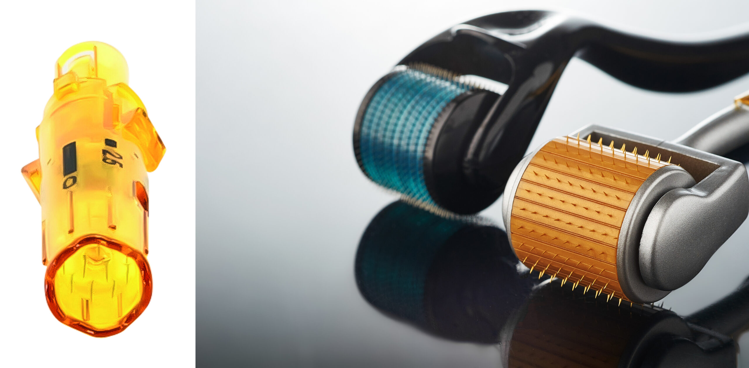
The two main methods of microneedling include using a pen and using a roller. Each of these categories includes many different products with a wide variety of options, settings, and needle lengths. Drs Davies and Miron recommend the Dermapen, a pen with disposable needles whose size can be adjusted as necessary during treatment. As they explain, “The Dermapen is an electrically powered medical device that delivers a vibrating stamplike motion to the skin, resulting in a series of microchannels.” Drs Garg and Rossi, on the other hand, recommend using a roller: “Because of the action of the roller, the needle penetrates the skin at an angle and then goes deeper as the roller turns.”

Microneedling Procedure

The microneedling procedure itself is actually quite simple, especially when compared with procedures utilizing dermal fillers or botulinum toxin, which require precise injections into very specific areas of the face. Topical anesthesia is applied to the face, and the clinician waits for the anesthesia to take effect. The specific application method of the microneedles will depend on the type of device chosen and the area of the face to be treated.

After the procedure, topical adjuncts such as PRP, PRF, or HA can be applied to the area. Because of the microchannels created in the skin, it is extremely receptive to the beneficial healing properties of these products at this time.

Side effects of the procedure are very mild. Any bleeding that occurs during the procedure should stop very quickly. There may be bruising and erythema that can last a few days. Depending on the severity of the patient’s condition, additional treatments may be required, but patients should wait 1 to 2 months before undergoing an additional microneedling procedure.

Comparison with At-Home Treatment

One unique aspect of microneedling is that it is a procedure that can be performed at home by patients themselves as well as by medical professionals. Of course, the two treatments are quite different. As Drs Garg and Rossi point out, “The key difference in these devices is the length of the needles. Most consumer models have needles of 0.2 to 0.3 mm in length. Medical-grade dermal needling devices have needles ranging from 0.2 mm up to 3.0 mm because they are designed to treat specific conditions.” Dermal rollers are available for purchase for as little as $10 or less, while medical-grade devices cost in the hundreds or thousands of dollars range. Clearly, one of these devices will be more effective than the other.

While the shorter needles of consumer microneedling products ensure that they carry less risk than with longer needles, the fact remains that the skin is being punctured by a user with less knowledge of facial anatomy, who may be using more or less force than is recommended, and who is likely not working with sterile equipment and in a sterile environment. In addition, at-home procedures may be more painful for the patient because they will be performed without anesthesia. Also, patients will not have access to adjuncts such as PRP, PRF, or HA to apply to the skin afterward. There are many topical products that consumers can use after an at-home microneedling procedure, but because these will go deeper into the skin than normal, it is necessary for patients to educate themselves on what products are safe and which may be harmful.

Conclusion

The more dentists who begin offering microneedling procedures as part of their practice, the more patients who can benefit.

Overall, while at-home treatment may be a good alternative for patients on a tight budget and who desire nothing more than a simple refresh of the skin, those who want more significant results will seek out professional treatment. The more dentists who begin offering microneedling procedures as part of their practice, the more patients who can benefit. Patients who have already been seeing you for years may feel more comfortable undergoing this treatment with someone they already trust rather than finding a new practitioner. Also, because even medical-grade microneedling is a simpler procedure to learn and perform than dermal fillers or botulinum toxin injections, it is a perfect “beginner” cosmetic procedure for clinicians considering expanding their practice. What better way is there to test the waters and find out whether including more cosmetic treatments would be right for you and your patients than to start with something small and simple? Microneedling is often used as an adjunct to dermal fillers, and dermal fillers may be used as an adjunct to Botox, so if you expand your practice further, your microneedling patients may become your first advanced cosmetic procedure patients.

Are you ready to take the next step? These new and forthcoming cosmetic dentistry titles are a great place to start:
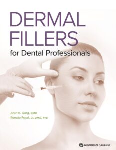
Dermal Fillers for Dental Professionals
Arun K. Garg and Renato Rossi, Jr

Esthetic medical procedures have evolved from the invasive, complex face-lift surgeries of the past to the modern practice of minimally invasive injections of dermal fillers, neurotoxins, and autologous agents. This book introduces these techniques and explains how to choose the most appropriate method for the situation, then provides precise instructions on how to administer different treatments. Photographic and illustrated diagrams explicitly demonstrate both the “where” and the “how” of the facial injection techniques, so clinicians can feel confident about applying these protocols and discovering the impressive results that will make patients feel most comfortable in their skin.
Coming soon! Join our mailing list for updates!
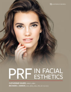
PRF in Facial Esthetics
Edited by Catherine Davies and Richard J. Miron

Written in collaboration with international experts from various fields of medicine, including basic scientists, clinician-scientists, experts in laser therapy and photography, as well as plastic surgeons and hair restorative surgeons, this book collectively offers a comprehensive approach to using platelet-rich fibrin (PRF) in facial esthetics. PRF has been used for decades in regenerative medicine, and slowly it has made its way into the medical esthetic arena, often used in combination with other leading therapies to support minimally invasive esthetic procedures. This book therefore starts at the beginning, first exploring the biology and anatomy of the skin and hair before turning to clinical chapters on patient consultation, microneedling, skin and hair regeneration, lasers, and the use of PRF in plastic surgery. The final chapter looks to the future and considers what else could be possible. If you perform any facial esthetic procedures in your office or want to learn how, this book is a must.

248 pp; 380 illus; ©2020; ISBN 978-0-86715-957-8 (B9578); $168
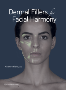
Dermal Fillers for Facial Harmony
Altamiro Flávio

In our esthetics-obsessed culture, patients are becoming more aware and demanding of dermal filler treatments. Yet all too often we hear news stories of botched procedures or anesthesia gone wrong. This book walks you through how to incorporate dermal fillers into your clinical armamentarium, starting with the basic characteristics of the materials and advancing to the injection techniques and clinical indications for their use. The author includes such fine detail as whether to use a needle versus a cannula and why you must understand the precise tissue layers of the face in order to effectively deliver this nuanced treatment. In step with its sister volume, Botulinum Toxin for Facial Harmony, the book focuses on facial analysis as a crucial step in determining appropriate treatment, a philosophy that is carried through the entire book. An entire chapter is devoted to clinical cases illustrating the various facial regions and their appropriate filler applications, clearly demonstrating how to use the techniques presented to optimize esthetics and facial harmony. Supplemental material and videos are available via QR code for additional learning.

176 pp; 228 illus; ©2019; ISBN 978-0-86715-821-2 (B8212); US $148
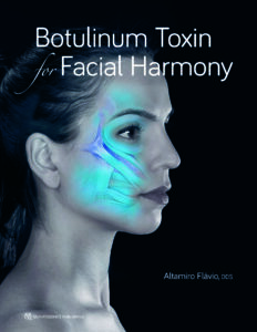
Botulinum Toxin for Facial Harmony
Altamiro Flávio

This book shows practitioners how to address facial asymmetries with clinical Botox therapy. The book covers comprehensive facial analysis and how to plan treatment. Injection points are identified within a larger discussion of anatomical considerations, and detailed treatment instructions include dosage, syringe type, and needle size for each clinical procedure as well as how to evaluate the results. This stunning book will widen the esthetic treatment options you can provide for patients.

160 pp; 359 illus; ©2018; ISBN 978-0-86715-787-1 (B7871); US $148

Posted in Books, Esthetic Dentistry, Misc, Multidisciplinary, Practice Management, What's New | Leave a comment

Probing depths? Check. Look for caries? Check. Additional screenings…?

Reading time: 9 minutes

Written by: Marieke Zaffron

What else do you screen your patients for during the dental examination? Do you have them fill out a questionnaire before their appointment? Or is that only for new patients? Do you find time to ask them questions about their health? Or do trust that the patient will tell you if something is wrong?

For many patients, they see you and your hygiene staff more often than any other medical professional. The patient may not realize that an annoying but nonpainful symptom such as TMJ clicking might be a sign of something more serious, and they may not bring it up unless prompted. No one is expecting the dentist to perform a comprehensive physical exam, but there are several key areas of the head, neck, and mouth where asking a few brief questions and taking a closer look could potentially make a world of difference to your patients’ health.

Tongue Screening

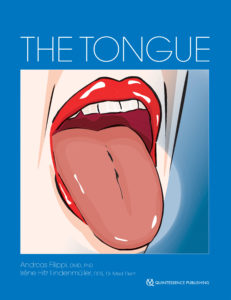
Let’s start in the oral cavity—we’re already there, right? How much attention are you giving to your patients’ tongues? According to Drs Andreas Filippi and Irène Hitz Lindenmüller, editors of The Tongue (Quintessence, 2019), “the borders, underside, and base of the tongue as well as the floor of the mouth should be inspected as a basic principle. If there are any visible or merely palpable changes, further diagnostic investigation should be discussed.” Ask the patient if they’ve had any reduced saliva flow or burning sensation of the tongue. Dozens of medical conditions—oral and general—present with signs and symptoms in the tongue, so it’s important to recognize any abnormalities that might be cause for concern. Even something as simple as a change in the color of the tongue might indicate problems.

A thorough tongue examination includes pulling the tongue out with a swab to allow the entire border of the tongue to be evaluated.

Oral Pathology Screening

A patient may have immaculate oral hygiene, no gingival recession or caries, and perfect teeth that need no management beyond the daily brushing and flossing. But that doesn’t mean they are free from oral diseases. As Dr Robert Marx, author of Oral Pathology in Clinical Practice (Quintessence, 2017) points out, “Dentists and their dental hygiene team historically have been the great identifiers of oral diseases—not only the common ones of dental caries and periodontal inflammation but also those of oral cancers, serious infections, systemic diseases, and many more.” In fact, taking notice of an unusual lesion and referring the patient to the correct specialist to identify it further can even save a life. Dr Marx recommends taking a close look at “the more hidden places in the oral cavity such as the posterior lingual mandible, the hamular notch area, and the posterior lateral border of the tongue … it is also prudent to examine the face and neck for obvious skin lesions and masses as well as the patient’s overall appearance and gait.” In addition, having a basic knowledge of oral pathology can help you to direct your patients to the correct specialist when there is a problem. In cases of certain cancers, time may be of the essence.

Squamous cell carcinoma that was incorrectly diagnosed and treated as gum disease for 2 years, giving the cancer time to invade the bone.

Orofacial Pain/TMD Screening

Now that we’ve covered the mouth, let’s move on to the rest of the head. Getting a correct orofacial pain diagnosis is especially important for dentists because of the prevalence of referred pain, or pain that is felt in one area even if the painful stimulus originates somewhere else. Your patients may be complaining of a toothache when the tooth itself is actually fine, but the patient has an undiagnosed pain disorder affecting the trigeminal nerve. Imagine if the patient was put through root canal treatment—or even had the tooth extracted—only for the pain to remain and the real problem to remain unresolved.

Drs Reny de Leeuw and Gary Klasser, editors of the sixth edition of Orofacial Pain: Guidelines for Assessment, Diagnosis, and Management (Quintessence, 2018), encourage dentists to screen all of their patients for TMDs and other orofacial pain disorders. The actual process of diagnosing a pain disorder can be involved, requiring a comprehensive history and clinical examination as well as diagnostic tests. However, adding a few simple screening questions to the dental exam is a great place to start. A positive response to any of these questions may be sufficient to warrant further investigation, including referral to a specialist.

Example screening questions for TMDs.

Sleep Apnea/Hypopnea Screening

Obstructive sleep apnea/hypopnea is a serious concern affecting 65 million Americans. Although sleep dentistry is developing as a specialty of its own, even general dentists should be aware of some of the basics to screen their patients. As Drs Gilles Lavigne, Peter Cistulli, and Michael Smith, editors of the new edition of Sleep Medicine for Dentists (Quintessence, 2020) put it, “It is generally accepted that the dentist’s role is to screen for potential sleep-disordered breathing (SDB) but not to diagnose SDB … Because patients with SDB may present to their dentist before their medical doctor, dentists have the potential to recognize patients with SDB who might not otherwise be diagnosed.” Both children and adults can suffer from SDB, so pediatric patients should be screened as well as older patients.

Screening for sleep disorders includes questioning patients on their sleep habits. Consider having your patients fill out a validated sleep questionnaire as part of their history forms. Check for the following symptoms of SDB.

Symptoms related to SDB.

Conclusion

Most patients don’t go to their dentists expecting to be diagnosed with a problem unrelated to their teeth. And of course, you probably won’t be the one doing the actual diagnosing for concerns that aren’t strictly dental. But even so, taking that extra step and being able to tell patients when they might have a problem, and having someone in your back pocket to refer them to so they can get the help they may need, can make all the difference.

Check out the titles below for more information on each of these topics.


The Tongue
Edited by Andreas Filippi and Irène Hitz Lindenmüller

As the largest organ in the oral cavity, the tongue not only plays a primary role in masticatory and speech function—it is also a significant indicator of health, demonstrating signs of both oral pathologies and diseases that can affect the entire body. Because no health care provider gets the opportunity to examine a patient’s tongue as often as the dentist, it is essential for dentists to recognize when there may be a problem with the tongue and what the problem is. In addition to an overview of tongue anatomy and general diagnosis and treatment recommendations, this book contains an atlas of more than 50 specific diseases and health concerns that may present signs and symptoms in the tongue. Each is outlined in a quick-reference table describing etiology, prognosis, and more and is accompanied by photographs of different ways the condition can present. A true diagnostic aid, this guide will allow clinicians to identify and address any abnormality a patient’s tongue may exhibit.

216 pp; 591 illus; ISBN: 978-0-86715-776-5; 9780867157765; US$148

Learn more here: What Dentists Should Know About Treating the Tongue

Oral Pathology in Clinical Practice
By Robert E. Marx

While most dentists do not perform their own histologic testing, all dentists must be able to recognize conditions that may require biopsy or further treatment outside the dentist office. This book does not pretend to be an exhaustive resource on oral pathology; instead, it seeks to provide the practicing clinician with enough information to help identify or at least narrow down the differential for every common lesion or oral manifestation of disease seen in daily practice as well as what to do about them. Organized by type of lesion, mass, or disease, each pathologic entity presented includes the nature of the disease; its predilections, clinical features, radiographic presentation, differential diagnosis, and microscopic features; and the suggested course of action for the dental practitioner as well as the standard treatment regimen. In keeping with the concise nature of the text, all but the rarest disease entities include at least one photograph to illustrate the clinical condition. This book distills the comprehensive information from Dr Marx and Dr Diane Stern’s award-winning pathology reference text (Oral and Maxillofacial Pathology: A Rationale for Diagnosis and Treatment, Second Edition [Quintessence, 2012]) into practical guidelines for restorative and general dentists everywhere.

376 pp; 425 illus; ISBN: 978-0-86715-764-2; 9780867157642; US$98

Learn more here: Clinical Pathology for Your Clinic

Orofacial Pain: Guidelines for Assessment, Diagnosis, and Management, Sixth Edition
Edited by Reny de Leeuw and Gary D. Klasser

Following in the tradition of the previous editions, this book offers the latest research and most up-to-date information on orofacial pain, including a concise overview of each condition as well as its symptoms, comorbidities, differential diagnosis, and treatment options. Every chapter has undergone critical updates to reflect the developments in the expanding field of orofacial pain, including the glossary. These updates include the addition of new diseases such as first-bite syndrome, revised information on genetic factors to reflect new insights gleaned from the OPPERA studies, expanded information on management strategies for certain conditions, and revisions to screening tools for biobehavioral factors. In addition, the classification system used in the fifth edition has been replaced and extended and includes DSM-5 codes where applicable. This book remains an essential tool for any health care provider who treats patients with orofacial pain.

336 pp; (softcover); 9 illus; ISBN: 978-0-86715-768-0; 9780867157680; US$56

Learn more here: Orofacial Pain Management in Dentistry

Sleep Medicine for Dentists: An Evidence-Based Overview, Second Edition
Edited by Gilles J. Lavigne, Peter A. Cistulli, and Michael T. Smith

Dentists are often the first medical practitioners to encounter patient reports or clinical evidence of disorders such as sleep apnea, sleep bruxism, and sleep-disrupting orofacial pain, providing them a unique opportunity to prevent the development or persistence of conditions that strongly impact their patients’ lives. Since the first publication of this seminal book, significant advances have been made in the field of sleep medicine, and this updated edition gathers all of this new evidence-based knowledge and presents it in focused, concise chapters. Leading experts in medicine and dentistry explain the neurobiologic mechanisms of sleep and how they can be affected by breathing disorders, bruxism, and pain, along the way guiding dental practitioners in performing their specific responsibilities for screening, treating, and often referring patients as part of a multidisciplinary team of physicians. An emphasis is placed on research findings regarding newly emerging cognitive behavioral approaches to treatment that mitigate some of the risks associated with pharmacologic and oral appliance therapies. Readers will find this book both fascinating and clinically important as they strive to provide the best possible treatment to patients with these complex and often life-threatening disorders.

224 pp; 40 illus ISBN: 978-0-86715-828-1; 9780867158281; US$78

Learn more here: Obstructive Sleep Apnea, the Didgeridoo, and You

The Clinician’s Handbook for Dental Sleep Medicine
Edited by Ken Berley and Steven Carstensen

It has been estimated that 20 million Americans suffer from moderate to severe OSA, and at least one patient in five has mild OSA. The primary treatment prescribed by sleep physicians is CPAP, but patient compliance with this therapy is unacceptably low, between 25% and 50%. There is a significant opportunity for dentists to provide a viable alternative therapy—oral appliance therapy (OAT). OAT results in much better adherence to therapy than CPAP, and while OAT is not as efficacious as CPAP, this increased compliance results in comparable therapeutic results. Currently, a board-certified sleep physician is the only medical professional qualified to diagnose OSA and other sleep-related breathing disorders (SRBDs), so dentists must coordinate with a sleep physician to provide OAT. This book is the how-to guide, a gateway to a successful dental sleep medicine practice. Written by two experts in the field, it clearly delineates the dentist’s role in the treatment of SRBDs and gives practical advice for how to incorporate dental sleep medicine into an existing dental practice, not to mention how to work with sleep physicians to best support patient care. In addition to step-by-step instructions for examination, appliance selection, and follow-up care, complications of OAT, legal issues, and medical insurance and Medicare considerations are included to fully prepare the dentist for the journey into dental sleep medicine.

240 pp; (softcover); 60 illus; ISBN: 978-0-86715-813-7; 9780867158137; US$72

Learn more here: Obstructive Sleep Apnea: A Deadly Disease with a Dental Solution

Posted in Books, Misc, Multidisciplinary, Occlusion & TMD, Sleep Dentistry | Leave a comment